Researchers in Australia have developed a nanosensor that can detect the onset of gestational diabetes with 95% accuracy. Demonstrated by a team led by Carlos Salomon at the University of Queensland, the superparamagnetic “nanoflower” sensor could enable doctors to detect a variety of complications in the early stages of pregnancy.
Many complications in pregnancy can have profound and lasting effects on both the mother and the developing foetus. Today, these conditions are detected using methods such as blood tests, ultrasound screening and blood pressure monitoring. In many cases, however, their sensitivity is severely limited in the earliest stages of pregnancy.
“Currently, most pregnancy complications cannot be identified until the second or third trimester, which means it can sometimes be too late for effective intervention,” Salomon explains.
To tackle this challenge, Salomon and his colleagues are investigating the use of specially engineered nanoparticles to isolate and detect biomarkers in the blood associated with complications in early pregnancy. Specifically, they aim to detect the protein molecules carried by extracellular vesicles (EVs) – tiny, membrane-bound particles released by the placenta, which play a crucial role in cell signalling.
In their previous research, the team pioneered the development of superparamagnetic nanostructures that selectively bind to specific EV biomarkers. Superparamagnetism occurs specifically in small, ferromagnetic nanoparticles, causing their magnetization to randomly flip direction under the influence of temperature. When proteins are bound to the surfaces of these nanostructures, their magnetic responses are altered detectably, providing the team with a reliable EV sensor.
“This technology has been developed using nanomaterials to detect biomarkers at low concentrations,” explains co-author Mostafa Masud. “This is what makes our technology more sensitive than current testing methods, and why it can pick up potential pregnancy complications much earlier.”
Previous versions of the sensor used porous nanocubes that efficiently captured EVs carrying a key placental protein named PLAP. By detecting unusual levels of PLAP in the blood of pregnant women, this approach enabled the researchers to detect complications far more easily than with existing techniques. However, the method generally required detection times lasting several hours, making it unsuitable for on-site screening.
In their latest study, reported in Science Advances, Salomon’s team started with a deeper analysis of the EV proteins carried by these blood samples. Through advanced computer modelling, they discovered that complications can be linked to changes in the relative abundance of PLAP and another placental protein, CD9.
Based on these findings, they developed a new superparamagnetic nanosensor capable of detecting both biomarkers simultaneously. Their design features flower-shaped nanostructures made of nickel ferrite, which were embedded into specialized testing strips to boost their sensitivity even further.
Using this sensor, the researchers collected blood samples from 201 pregnant women at 11 to 13 weeks’ gestation. “We detected possible complications, such as preterm birth, gestational diabetes and preeclampsia, which is high blood pressure during pregnancy,” Salomon describes. For gestational diabetes, the sensor demonstrated 95% sensitivity in identifying at-risk cases, and 100% specificity in ruling out healthy cases.
Based on these results, the researchers are hopeful that further refinements to their nanoflower sensor could lead to a new generation of EV protein detectors, enabling the early diagnosis of a wide range of pregnancy complications.
“With this technology, pregnant women will be able to seek medical intervention much earlier,” Salomon says. “This has the potential to revolutionize risk assessment and improve clinical decision-making in obstetric care.”
When a mantis shrimp uses shock waves to strike and kill its prey, how does it prevent those shock waves from damaging its own tissues? Researchers at Northwestern University in the US have answered this question by identifying a structure within the shrimp that filters out harmful frequencies. Their findings, which they obtained by using ultrasonic techniques to investigate surface and bulk wave propagation in the shrimp’s dactyl club, could lead to novel advanced protective materials for military and civilian applications.
Dactyl clubs are hammer-like structures located on each side of a mantis shrimp’s body. They store energy in elastic structures similar to springs that are latched in place by tendons. When the shrimp contracts its muscles, the latch releases, releasing the stored energy and propelling the club forward with a peak force of up to 1500 N.
This huge force (relative to the animal’s size) creates stress waves in both the shrimp’s target – typically a hard-shelled animal such as a crab or mollusc – and the dactyl club itself, explains biomechanical engineer Horacio Dante Espinosa, who led the Northwestern research effort. The club’s punch also creates bubbles that rapidly collapse to produce shockwaves in the megahertz range. “The collapse of these bubbles (a process known as cavitation collapse), which takes place in just nanoseconds, releases intense bursts of energy that travel through the target and shrimp’s club,” he explains. “This secondary shockwave effect makes the shrimp’s strike even more devastating.”
Protective phononic armour
So how do the shrimp’s own soft tissues escape damage? To answer this question, Espinosa and colleagues studied the animal’s armour using transient grating spectroscopy (TGS) and asynchronous optical sampling (ASOPS). These ultrasonic techniques respectively analyse how stress waves propagate through a material and characterize the material’s microstructure. In this work, Espinosa and colleagues used them to provide high-resolution, frequency-dependent wave propagation characteristics that previous studies had not investigated experimentally.
The team identified three distinct regions in the shrimp’s dactyl club. The outermost layer consists of a hard hydroxyapatite coating approximately 70 μm thick, which is durable and resists damage. Beneath this, an approximately 500 μm-thick layer of mineralized chitin fibres arranged in a herringbone pattern enhances the club’s fracture resistance. Deeper still, Espinosa explains, is a region that features twisted fibre bundles organized in a corkscrew-like arrangement known as a Bouligand structure. Within this structure, each successive layer is rotated relative to its neighbours, giving it a unique and crucial role in controlling how stress waves propagate through the shrimp.
“Our key finding was the existence of phononic bandgaps (through which waves within a specific frequency range cannot travel) in the Bouligand structure,” Espinosa explains. “These bandgaps filter out harmful stress waves so that they do not propagate back into the shrimp’s club and body. They thus preserve the club’s integrity and protect soft tissue in the animal’s appendage.”
The team also employed finite element simulations incorporating so-called Bloch-Floquet analyses and graded mechanical properties to understand the phonon bandgap effects. The most surprising result, Espinosa tells Physics World, was the formation of a flat branch around the 450 to 480 MHz range, which correlates to frequencies arising from bubble collapse originating during club impact.
Evolution and its applications
For Espinosa and his colleagues, a key goal of their research is to understand how evolution leads to natural composite materials with unique photonic, mechanical and thermal properties. In particular, they seek to uncover how hierarchical structures in natural materials and the chemistry of their constituents produce emergent mechanical properties. “The mantis shrimp’s dactyl club is an example of how evolution leads to materials capable of resisting extreme conditions,” Espinosa says. “In this case, it is the violent impacts the animal uses for predation or protection.”
The properties of the natural “phononic shield” unearthed in this work might inspire advanced protective materials for both military and civilian applications, he says. Examples could include the design of helmets, personnel armour, and packaging for electronics and other sensitive devices.
In this study, which is described in Science, the researchers analysed two-dimensional simulations of wave behaviour. Future research, they say, should focus on more complex three-dimensional simulations to fully capture how the club’s structure interacts with shock waves. “Designing aquatic experiments with state-of-the-art instrumentation would also allow us to investigate how phononic properties function in submerged underwater conditions,” says Espinosa.
The team would also like to use biomimetics to make synthetic metamaterials based on the insights gleaned from this work.
Physicists in Austria have shown that the static electricity acquired by identical material samples can evolve differently over time, based on each samples’ history of contact with other samples. Led by Juan Carlos Sobarzo and Scott Waitukaitis at the Institute of Science and Technology Austria, the team hope that their experimental results could provide new insights into one of the oldest mysteries in physics.
Static electricity – also known as contact electrification or triboelectrification — has been studied for centuries. However, physicists still do not understand some aspects of how it works.
“It’s a seemingly simple effect,” Sobarzo explains. “Take two materials, make them touch and separate them, and they will have exchanged electric charge. Yet, the experiments are plagued by unpredictability.”
This mystery is epitomized by an early experiment carried out by the German-Swedish physicist Johan Wilcke in 1757. When glass was touched to paper, Wilcke found that glass gained a positive charge – while when paper was touched to sulphur, it would itself become positively charged.
Triboelectric series
Wilcke concluded that glass will become positively charged when touched to sulphur. This concept formed the basis of the triboelectric series, which ranks materials according to the charge they acquire when touched to another material.
Yet in the intervening centuries, the triboelectric series has proven to be notoriously inconsistent. Despite our vastly improved knowledge of material properties since the time of Wilcke’s experiments, even the latest attempts at ordering materials into triboelectric series have repeatedly failed to hold up to experimental scrutiny.
According to Sobarzo’s and colleagues, this problem has been confounded by the diverse array of variables associated with a material’s contact electrification. These include its electronic properties, pH, hydrophobicity, and mechanochemistry, to name just a few.
In their new study, the team approached the problem from a new perspective. “In order to reduce the number of variables, we decided to use identical materials,” Sobarzo describes. “Our samples are made of a soft polymer (PDMS) that I fabricate myself in the lab, cut from a single piece of material.”
Starting from scratch
For these identical materials, the team proposed that triboelectric properties could evolve over time as the samples were brought into contact with other, initially identical samples. If this were the case, it would allow the team to build a triboelectric series from scratch.
At first, the results seemed as unpredictable as ever. However, as the same set of samples underwent repeated contacts, the team found that their charging behaviour became more consistent, gradually forming a clear triboelectric series.
Initially, the researchers attempted to uncover correlations between this evolution and variations in the parameters of each sample – with no conclusive results. This led them to consider whether the triboelectric behaviour of each sample was affected by the act of contact itself.
Contact history
“Once we started to keep track of the contact history of our samples – that is, the number of times each sample has been contacted to others–the unpredictability we saw initially started to make sense,” Sobarzo explains. “The more contacts samples would have in their history, the more predictable they would behave. Not only that, but a sample with more contacts in its history will consistently charge negative against a sample with less contacts in its history.”
To explain the origins of this history-dependent behaviour, the team used a variety of techniques to analyse differences between the surfaces of uncontacted samples, and those which had already been contacted several times. Their measurements revealed just one difference between samples at different positions on the triboelectric series. This was their nanoscale surface roughness, which smoothed out as the samples experienced more contacts.
“I think the main take away is the importance of contact history and how it can subvert the widespread unpredictability observed in tribocharging,” Sobarzo says. “Contact is necessary for the effect to happen, it’s part of the name ‘contact electrification’, and yet it’s been widely overlooked.”
The team is still uncertain of how surface roughness could be affecting their samples’ place within the triboelectric series. However, their results could now provide the first steps towards a comprehensive model that can predict a material’s triboelectric properties based on its contact-induced surface roughness.
Sobarzo and colleagues are hopeful that such a model could enable robust methods for predicting the charges which any given pair of materials will acquire as they touch each other and separate. In turn, it may finally help to provide a solution to one of the most long-standing mysteries in physics.
The COVID-19 pandemic provided a driving force for researchers to seek out new disinfection methods that could tackle future viral outbreaks. One promising approach relies on the use of nanoparticles, with several metal and metal oxide nanoparticles showing anti-viral activity against SARS-CoV-2, the virus that causes COVID-19. With this in mind, researchers from Sweden and Estonia investigated the effect of such nanoparticles on two different virus types.
Aiming to elucidate the nanoparticles’ mode of action, they discovered a previously unknown antiviral mechanism, reporting their findings in Nanoscale.
The researchers – from the Swedish University of Agricultural Sciences (SLU) and the University of Tartu – examined triethanolamine terminated titania (TATT) nanoparticles, spherical 3.5-nm diameter titanium dioxide (titania) particles that are expected to interact strongly with viral surface proteins.
They tested the antiviral activity of the TATT nanoparticles against two types of virus: swine transmissible gastroenteritis virus (TGEV) – an enveloped coronavirus that’s surrounded by a phospholipid membrane and transmembrane proteins; and the non-enveloped encephalomyocarditis virus (EMCV), which does not have a phospholipid membrane. SARS-CoV-2 has a similar structure to TGEV: an enveloped virus with an outer lipid membrane and three proteins forming the surface.
In this latest investigation, the team aimed to determine whether one of these potential mechanisms – blocking of surface proteins, or membrane disruption via oxidation by nanoparticle-generated reactive oxygen species – is the likely cause of TATT’s antiviral activity. The first of these effects usually occurs at low (nanomolar to micromolar) nanoparticle concentrations, the latter at higher (millimolar) concentrations.
Mode of action
To assess the nanoparticle’s antiviral activity, the researchers exposed viral suspensions to colloidal TATT solutions for 1 h, at room temperature and in the dark (without UV illumination). For comparison, they repeated the process with silicotungstate polyoxometalate (POM) nanoparticles, which are not able to bind strongly to cell membranes.
The nanoparticle-exposed viruses were then used to infect cells and the resulting cell viability served as a measure of the virus infectivity. The team note that the nanoparticles alone showed no cytotoxicity against the host cells.
Measuring viral infectivity after nanoparticle exposure revealed that POM nanoparticles did not exhibit antiviral effects on either virus, even at relatively high concentrations of 1.25 mM. TATT nanoparticles, on the other hand, showed significant antiviral activity against the enveloped TGEV virus at concentrations starting from 0.125 mM, but did not affect the non-enveloped EMCV virus.
Based on previous evidence that TATT nanoparticles interact strongly with proteins in darkness, the researchers expected to see antiviral activity at a nanomolar level. But the finding that TATT activity only occurred at millimolar concentrations, and only affected the enveloped virus, suggests that the antiviral effect is not due to blocking of surface proteins. And as titania is not oxidative in darkness, the team propose that the antiviral effect is actually due to direct complexation of nanoparticles with membrane phospholipids – a mode of antiviral action not previously considered.
“Typical nanoparticle concentrations required for effects on membrane proteins correspond to the protein content on the virus surface. With a 1:1 complex, we would need maximum nanomolar concentrations,” Kessler explains. “We saw an effect at about 1 mM/l, which is far higher. This was the indication for us that the effect was on the whole of membrane.”
Verifying the membrane effect
To corroborate their hypothesis, the researchers examined the leakage of dye-labelled RNA from the TGEV coronavirus after 1 h exposure to nanoparticles. The fluorescence signal from the dye showed that TATT-treated TGEV released significantly more RNA than non-exposed virus, attributed to the nanoparticles disrupting the virus’s phospholipid membrane.
Finally, the team studied the interactions between TATT nanoparticles and two model phospholipid compounds. Both molecules formed strong complexes with TATT nanoparticles, while their interaction with POM nanoparticles was weak. This additional verification led the researchers to conclude that the antiviral effect of TATT in dark conditions is due to direct membrane disruption via complexation of titania nanoparticles with phospholipids.
“To the best of our knowledge, [this] proves a new pathway for metal oxide nanoparticles antiviral action,” they write.
Importantly, the nanoparticles are non-toxic, and work at room temperature without requiring UV illumination – enabling simple and low-cost disinfection methods. “While it was known that disinfection with titania could work in UV light, we showed that no special technical measures are necessary,” says Kessler.
Kessler suggests that the nanoparticles could be used to coat surfaces to destroy enveloped viruses, or in cost-effective filters to decontaminate air or water. “[It should be] possible to easily create antiviral surfaces that don’t require any UV activation just by spraying them with a solution of TATT, or possibly other oxide nanoparticles with an affinity to phosphate, including iron and aluminium oxides in particular,” he tells Physics World.
A new type of quantum bit (qubit) that stores information in a quantum dot with the help of an ensemble of nuclear spin states has been unveiled by physicists in the UK and Austria. Led by Dorian Gangloff and Mete Atatüre at the University of Cambridge, the team created a collective quantum state that could be used as a quantum register to store and relay information in a quantum communication network of the future.
Quantum communication networks are used to exchange and distribute quantum information between remotely-located quantum computers and other devices. As well as enabling distributed quantum computing, quantum networks can also support secure quantum cryptography. Today, these networks are in the very early stages of development and use the entangled quantum states of photons to transmit information. Network performance is severely limited by decoherence, whereby the quantum information held by photons is degraded as they travel long distances. As a result, effective networks need repeater nodes that receive and then amplify weakened quantum signals.
“To address these limitations, researchers have focused on developing quantum memories capable of reliably storing entangled states to enable quantum repeater operations over extended distances,” Gangloff explains. “Various quantum systems are being explored, with semiconductor quantum dots being the best single-photon generators delivering both photon coherence and brightness.”
Single-photon emission
Quantum dots are widely used for their ability to emit single photons at specific wavelengths. These photons are created by electronic transitions in quantum dots and are ideal for encoding and transmitting quantum information.
However, the electronic spin states of quantum dots are not particularly good at storing quantum information for long enough to be useful as stationary qubits (or nodes) in a quantum network. This is because they contain hundreds or thousands of nuclei with spins that fluctuate. The noise generated by these fluctuations causes the decoherence of qubits based on electronic spin states.
In their previous research, Gangloff and Atatüre’s team showed how this noise could be controlled by sensing how it interacts with the electronic spin states.
Atatüre says, “Building on our previous achievements, we suppressed random fluctuations in the nuclear ensemble using a quantum feedback algorithm. This is already very useful as it dramatically improves the electron spin qubit performance.”
Magnon excitation
Now, using a gallium arsenide quantum dot, the team has used the feedback algorithm to stabilize 13,000 nuclear spin states in a collective, entangled “dark state”. This is a stable quantum state that cannot absorb or emit photons. By introducing just a single nuclear magnon (spin flip) excitation, shared across all 13,000 nuclei, they could then flip the entire ensemble between two different collective quantum states.
Each of these collective states could respectively be defined as a 0 and a 1 in a binary quantum logic system. The team then showed how quantum information could be exchanged between the nuclear system and the quantum dot’s electronic qubit with a fidelity of about 70%.
“The quantum memory maintained the stored state for approximately 130 µs, validating the effectiveness of our protocol,” Gangloff explains. “We also identified unambiguously the factors limiting the current fidelity and storage time, including crosstalk between nuclear modes and optically induced spin relaxation.”
The researchers are hopeful that their approach could transform one of the biggest limitations to quantum dot-based communication networks into a significant advantage.
“By integrating a multi-qubit register with quantum dots – the brightest and already commercially available single-photon sources – we elevate these devices to a much higher technology readiness level,” Atatüre explains.
With some further improvements to their system’s fidelity, the researchers are now confident that it could be used to strengthen interactions between quantum dot qubits and the photonic states they produce, ultimately leading to longer coherence times in quantum communication networks. Elsewhere, it could even be used to explore new quantum phenomena, and gather new insights into the intricate dynamics of quantum many-body systems.
A newly-discovered class of quasiparticles known as fractional excitons offers fresh opportunities for condensed-matter research and could reveal unprecedented quantum phases, say physicists at Brown University in the US. The new quasiparticles, which are neither bosons nor fermions and carry no charge, could have applications in quantum computing and sensing, they say.
In our everyday, three-dimensional world, particles are classified as either fermions or bosons. Fermions such as electrons follow the Pauli exclusion principle, which prevents them from occupying the same quantum state. This property underpins phenomena like the structure of atoms and the behaviour of metals and insulators. Bosons, on the other hand, can occupy the same state, allowing for effects like superconductivity and superfluidity.
Fractional excitons defy this traditional classification, says Jia Leo Li, who led the research. Their properties lie somewhere in between those of fermions and bosons, making them more akin to anyons, which are particles that exist only in two-dimensional systems. But that’s only one aspect of their unusual nature, Li adds. “Unlike typical anyons, which carry a fractional charge of an electron, fractional excitons are neutral particles, representing a distinct type of quantum entity,” he says.
The experiment
Li and colleagues created the fractional excitons using two sheets of graphene – a form of carbon just one atom thick – separated by a layer of another two-dimensional material, hexagonal boron nitride. This layered setup allowed them to precisely control the movement of electrons and positively-charged “holes” and thus to generate excitons, which are pairs of electrons and holes that behave like single particles.
The team then applied a 12 T magnetic field to their bilayer structure. This strong field caused the electrons in the graphene to split into fractional charges – a well-known phenomenon that occurs in the fractional quantum Hall effect. “Here, strong magnetic fields create Landau electronic levels that induce particles with fractional charges,” Li explains. “The bilayer structure facilitates pairing between these positive and negative charges, making fractional excitons possible.”
“Distinct from any known particles”
The fractional excitons represent a quantum system of neutral particles that obey fractional quantum statistics, interact via dipolar forces and are distinct from any known particles, Li tells Physics World. He adds that his team’s study, which is detailed in Nature, builds on prior works that predicted the existence of excitons in the fractional quantum Hall effect (see, for example, Nature Physics 13, 751 2017, Nature Physics 15, 898-903 2019, Science 375 (6577), 205-209 2022).
The researchers now plan to explore the properties of fractional excitons further. “Our key objectives include measuring the fractional charge of the constituent particles and confirming their anyonic statistics,” Li explains. Studies of this nature could shed light on how fractional excitons interact and flow, potentially revealing new quantum phases, he adds.
“Such insights could have profound implications for quantum technologies, including ultra-sensitive sensors and robust quantum computing platforms,” Li says. “As research progresses, fractional excitons may redefine the boundaries of condensed-matter physics and applied quantum science.”
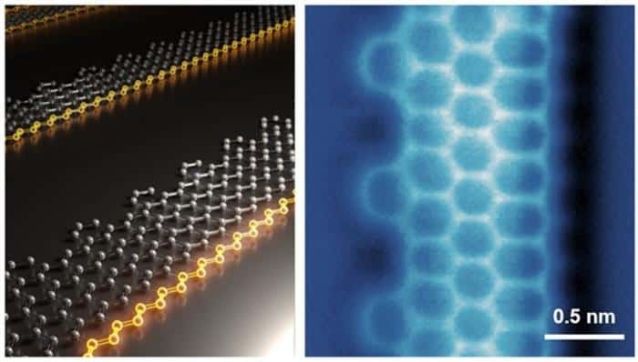
A new graphene nanostructure could become the basis for the first ferromagnets made purely from carbon. Known as an asymmetric or “Janus” graphene nanoribbon after the two-faced god in Roman mythology, the opposite edges of this structure have different properties, with one edge taking a zigzag form. Lu Jiong , a researcher at the National University of Singapore (NUS) who co-led the effort to make the structure, explains that it is this zigzag edge that gives rise to the ferromagnetic state, making the structure the first of its kind.
“The work is the first demonstration of the concept of a Janus graphene nanoribbon (JGNR) strand featuring a single ferromagnetic zigzag edge,” Lu says.
Graphene nanostructures with zigzag-shaped edges show much promise for technological applications thanks to their electronic and magnetic properties. Zigzag GNRs (ZGNRs) are especially appealing because the behaviour of their electrons can be tuned from metal-like to semiconducting by adjusting the length or width of the ribbons; modifying the structure of their edges; or doping them with non-carbon atoms. The same techniques can also be used to make such materials magnetic. This versatility means they can be used as building blocks for numerous applications, including quantum and spintronics technologies.
“It has been a long-sought goal to make other forms of zigzag-edge related GNRs with exotic quantum magnetic states for studying new science and developing new applications,” says team member Song Shaotang, the first author of a paper in Nature about the research.
ZGNRs with asymmetric edges
Building on topological classification theory developed in previous research by Louie and colleagues, theorists in the Singapore-Japan-US collaboration predicted that it should be possible to tune the magnetic properties of these structures by making ZGNRs with asymmetric edges. “These nanoribbons have one pristine zigzag edge and another edge decorated with a pattern of topological defects spaced by a certain number m of missing motifs,” Louie explains. “Our experimental team members, using innovative z-shaped precursor molecules for synthesis, were able to make two kinds of such ZGNRs. Both of these have one edge that supports a benzene motif array with a spacing of m = 2 missing benzene rings in between. The other edge is a conventional zigzag edge.”
Crucially, the theory predicted that the magnetic behaviour – ranging from antiferromagnetism to ferrimagnetism to ferromagnetism – of these JGNRs could be controlled by varying the value of m. In particular, says Louie, the configuration of m = 2 is predicted to show ferromagnetism – that is, all electron spins aligned in the same direction – concentrated entirely on the pristine zigzag edge. This behaviour contrasts sharply with that of symmetric ZGNRs, where spin polarization occurs on both edges and the aligned edge spins are antiferromagnetically coupled across the width of the ribbon.
Precursor design and synthesis
To validate these theoretical predictions, the team synthesized JGNRs on a surface. They then used advanced scanning tunnelling microscope (STM) and atomic force microscope (AFM) measurements to visualize the materials’ exact real-space chemical structure. These measurements also revealed the emergence of exotic magnetic states in the JGNRs synthesized in Lu’s lab at the NUS.
Two sides: An atomic model of the Janus graphene nanoribbons (left) and its atomic force microscopic image (right). (Courtesy: National University of Singapore)
In the past, Sakaguchi explains that GNRs were mainly synthesized using symmetric precursor chemical structures, largely because their asymmetric counterparts were so scarce. One of the challenges in this work, he notes, was to design asymmetric polymeric precursors that could undergo the essential fusion (dehydrogenation) process to form JGNRs. These molecules often orient randomly, so the researchers needed to use additional techniques to align them unidirectionally prior to the polymerization reaction. “Addressing this challenge in the future could allow us to produce JGNRs with a broader range of magnetic properties,” Sakaguchi says.
Towards carbon-based ferromagnets
According to Lu, the team’s research shows that JGNRs could become the first carbon-based spin transport channels to show ferromagnetism. They might even lead to the development of carbon-based ferromagnets, capping off a research effort that began in the 1980s.
However, Lu acknowledges that there is much work to do before these structures find real-world applications. For one, they are not currently very robust when exposed to air. “The next goal,” he says, “is to develop chemical modifications that will enhance the stability of these 1D structures so that they can survive under ambient conditions.”
A further goal, he continues, is to synthesize JGNRs with different values of m, as well as other classes of JGNRs with different types of defective edges. “We will also be exploring the 1D spin physics of these structures and [will] investigate their spin dynamics using techniques such as scanning tunnelling microscopy combined with electron spin resonance, paving the way for their potential applications in quantum technologies.”
Researchers at the University of Chicago’s Pritzker School of Molecular Engineering have created a groundbreaking hydrogel that doubles as a semiconductor. The material combines the soft, flexible properties of biological tissues with the electronic capabilities of semiconductors, making it ideal for advanced medical devices.
In a study published in Science, the research team, led by Sihong Wang, developed a stretchy, jelly-like material that provides the robust semiconducting properties necessary for use in devices such as pacemakers, biosensors and drug delivery systems.
Rethinking hydrogel design
Hydrogels are ideal for many biomedical applications because they are soft, flexible and water-absorbent – just like human tissues. Material scientists, long recognizing the vast potential of hydrogels, have pushed the boundaries of this class of material. One way is to create hydrogels with semiconducting abilities that can be useful for transmitting information between living tissues and bioelectronic device interfaces – in other words, a hydrogel semiconductor.
Imparting semiconducting properties to hydrogels is no easy task, however. Semiconductors, while known for their remarkable electronic properties, are typically rigid, brittle and water-repellent, making them inherently incompatible with hydrogels. By overcoming this fundamental mismatch, Wang and his team have created a material that could revolutionize the way medical devices interface with the human body.
Traditional hydrogels are made by dissolving hydrogel precursors (monomers or polymers) in water and adding chemicals to crosslink the polymers and form a water-swelled state. Since most polymers are inherently insulating, creating a hydrogel with semiconducting properties requires a special class of semiconducting polymers. The challenges do not stop there, however. These polymers typically only dissolve in organic solvents, not in water.
“The question becomes how to achieve a well-dispersed distribution of these semiconducting materials within a hydrogel matrix,” says first author Yahao Dai, a PhD student in the Wang lab. “This isn’t just about randomly dispersing particles into the matrix. To achieve strong electrical performance, a 3D interconnected network is essential for effective charge transport. So, the fundamental question is: how do you build a hydrophobic, 3D interconnected network within the hydrogel matrix?”
Innovative material Sihong Wang (left), Yahao Dai (right) and colleagues have developed a novel hydrogel with semiconducting properties. (Courtesy: UChicago Pritzker School of Molecular Engineering/John Zich)
To address this challenge, the researchers first dissolved the polymer in an organic solvent that is miscible with water, forming an organogel – a gel-like material composed of an organic liquid phase in a 3D gel network. They then immersed the organogel in water and allowed the water to gradually replace the organic solvent, transforming it into a hydrogel.
The researchers point out that this versatile solvent exchange process can be adapted to a variety of semiconducting polymers, opening up new possibilities for hydrogel semiconductors with diverse applications.
A two-in-one material
The result is a hydrogel semiconductor material that’s soft enough to match the feel of human tissue. With a Young’s modulus as low as 81 kPa – comparable to that of jelly – and the ability to stretch up to 150% of its original length, this material mimics the flexibility and softness of living tissue. These tissue-like characteristics allow the material to seamlessly interface with the human body, reducing the inflammation and immune responses that are often triggered by rigid medical implants.
The material also has a high charge carrier mobility, a measure of its ability to efficiently transmit electrical signals, of up to 1.4 cm2/V/s. This makes it suitable for biomedical devices that require effective semiconducting performance.
The potential applications extend beyond implanted devices. The material’s high hydration and porosity enable efficient volumetric biosensing and mass transport throughout the entire thickness of the semiconducting layer, which is useful for biosensing, tissue engineering and drug delivery applications. The hydrogel also responds to light effectively, opening up possibilities for light-controlled therapies, such as light-activated wireless pacemakers or wound dressings that use heat to accelerate healing.
A vision for transforming healthcare
The research team’s hydrogel material is now patented and being commercialized through UChicago’s Polsky Center for Entrepreneurship and Innovation. “Our goal is to further develop this material system and enhance its performance and application space,” says Dai. While the immediate focus is on enhancing the electrical and light modulation properties of the hydrogel, the team envisions future work in biochemical sensing.
“An important consideration is how to functionalize various bioreceptors within the hydrogel semiconductor,” explains Dai. “As each biomarker requires a specific bioreceptor, the goal is to target as many biomarkers as possible.”
The team is already exploring new methods to incorporate bioreceptors, such as antibodies and aptamers, within the hydrogels. With these advances, this class of semiconductor hydrogels could act as next-generation interfaces between human tissues and bioelectronic devices, from sensors to tailored drug-delivery systems. This breakthrough material may soon bridge the gap between living systems and electronics in ways once thought impossible.
Hot material The crystal structure of cordierite gives the material its unique thermal properties. (Courtesy: M Dove and L Li/Matter)
The anomalous and ultra-low thermal expansion of cordierite results from the interplay between lattice vibrations and the elastic properties of the material. That is the conclusion of Martin Dove at China’s Sichuan University and Queen Mary University of London in the UK and Li Li at the Civil Aviation Flight University of China. They showed that the material’s unusual behaviour stems from direction-varying elastic forces in its lattice, which act to vary cordierite’s thermal expansion along different directions.
Cordierite is a naturally-occurring mineral that can also be synthesized. Thanks to its remarkable thermal properties, it is used in products ranging from pizza stones to catalytic converters. When heated to high temperatures, it undergoes ultra-low thermal expansion along two directions, and it shrinks a tiny amount along the third direction. This makes it incredibly useful as a material that can be heated and cooled without changing size or suffering damage.
Despite its widespread use, scientists lack a fundamental understanding of how cordierite’s anomalous thermal expansion arises from the properties of its crystal lattice. Normally, thermal expansion (positive or negative) is understood in terms of Grüneisen parameters. These describe how vibrational modes (phonons) in the lattice cause it to expand or contract along each axis as the temperature changes.
Negative Grüneisen parameters describe a lattice that shrinks when heated, and are seen as key to understanding thermal contraction of cordierite. However, the material’s thermal response is not isotropic (it only contracts only along one axis when heated at high temperatures) so understanding cordierite in terms of its Grüneisen parameters alone is difficult.
Advanced molecular dynamics
In their study, Dove and Li used advanced molecular dynamics simulations to accurately model the behaviour of atoms in the cordierite lattice. Their closely matched experimental observations of the material’s thermal expansion, providing them with key insights into why the material has a negative thermal expansion in just one direction.
“Our research demonstrates that the anomalous thermal expansion of cordierite originates from a surprising interplay between atomic vibrations and elasticity,” Dove explains. The elasticity is described in the form of an elastic compliance tensor, which predicts how a material will distort in response to a force applied along a specific direction.
At lower temperatures, lattice vibrations occur at lower frequencies. In this case, the simulations predicted negative thermal expansion in all directions – which is in line with observations of the material.
At higher temperatures, the lattice becomes dominated by high-frequency vibrations. In principle, this should result in positive thermal expansion in all three directions. Crucially, however, Dove and Li discovered that this expansion is cancelled out by the material’s elastic properties, as described by its elastic compliance tensor.
What is more, the unique arrangement of crystal lattice meant that this tensor varied depending on the direction of the applied force, creating an imbalance that amplifies differences between the material’s expansion along each axis.
Cancellation mechanism
“This cancellation mechanism explains why cordierite exhibits small positive expansion in two directions and small negative expansion in the third,” Dove explains. “Initially, I was sceptical of the results. The initial data suggested uniform expansion behaviour at both high and low temperatures, but the final results revealed a delicate balance of forces. It was a moment of scientific serendipity.”
Altogether, Dove and Li’s result clearly shows that cordierite’s anomalous behaviour cannot be understood by focusing solely on the Grüneisen parameters of its three axes. It is crucial to take its elastic compliance tensor into account.
In solving this long-standing mystery, the duo now hope their results could help researchers to better predict how cordierite’s thermal expansion will vary at different temperatures. In turn, they could help to extend the useful applications of the material even further.
“Anisotropic materials like cordierite hold immense potential for developing high-performance materials with unique thermal behaviours,” Dove says. “Our approach can rapidly predict these properties, significantly reducing the reliance on expensive and time-consuming experimental procedures.”
Oil spills can pollute large volumes of surrounding water – thousands of times greater than the spill itself – causing long-term economic, environmental, social and ecological damage. Effective methods for in situ capture of spilled oil are thus essential to minimize contamination from such disasters.
Many oil spill cleanup technologies, however, exhibit poor hydrodynamic stability under complex flow conditions, which leads to poor oil-capture efficiency. To address this shortfall, researchers from Harbin Institute of Technology in China have come up with a new approach to oil cleanup using a vortex-anchored filter (VAF).
“Since the 1979 Atlantic Empress disaster, interception and adsorption have been the primary methods for oil spill recovery, but these are sensitive to water-flow fluctuation,” explains lead author Shijie You. Oil-in-water emulsions from leaking pipelines and offshore industrial discharge are particularly challenging, says You, adding that “these problems inspire us to consider how we can address hydrodynamic stability of oil-capture devices under turbulent conditions”.
Inspired by the natural world
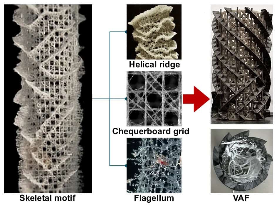
You and colleagues believe that the answers to oil spill challenges could come from nature – arguably the world’s greatest scientist. They found that the deep-sea glass sponge E. aspergillum, which lives at depths of up to 1000 m in the Pacific Ocean, has an excellent ability to filter feed with a high effectiveness, selectivity and robustness, and that its food particles share similarities with oil droplets.
The anatomical structure of E. aspergillum – also known as Venus’ flower basket – provided inspiration for the researchers to design their VAF. By mimicking the skeletal architecture and filter feeding patterns of the sponge, they created a filter that exhibited a high mass transfer and hydrodynamic stability in cleaning up oil spills under turbulent flow.
“The E. aspergillum has a multilayered skeleton–flagellum architecture, which creates 3D streamlines with frequent collision, deflection, convergence and separation,” explains You. “This can dissipate macro-scale turbulent flows into small-scale swirling flow patterns called low-speed vortical flows within the body cavity, which reduces hydrodynamic load and enhances interfacial mass transfer.”
For the sponges, this allows them to maintain a high mechanical stability while absorbing nutrients from the water. The same principles can be applied to synthetic materials for cleaning up oil spills.
VAF design Skeletal motif of E. aspergillum and (right column) front and top views of the VAF with a bio-inspired hollow cylinder skeleton and flagellum adsorbent. (Courtesy: Y Yu et al. Nat. Commun. 10.1038/s41467-024-55587-y)
The VAF is a synthetic form of the sponge’s architecture and, according to You, “is capable of transferring kinematic energy from an external water flow into multiple small-scale low-speed vortical flows within the body cavity to enhance hydrodynamic stability and oil capture efficiency”.
The tubular outer skeleton of the VAF comprises a helical ridge and chequerboard lattice. It is this skeleton that creates a slow vortex field inside the cavity and enables mass transfer of oil during the filtering process. Once the oil has been forced into the filter, the internal area – composed of flagellum-shaped adsorbent materials – provides a large interfacial area for oil adsorption.
Using the VAF to clean up oil spills
The researchers used their nature-inspired VAF to clean up oil spills under complex hydrodynamic conditions. You states that “the VAF can retain the external turbulent-flow kinetic energy in the low-speed vortical flows – with a small Kolmogorov microscale (85 µm) [the size of the smallest eddy in a turbulent flow] – inside the cavity of the skeleton, leading to enhanced interfacial mass transfer and residence time”.
“This led to an improvement in the hydrodynamic stability of the filter compared to other approaches by reducing the Reynolds stresses in nearly quiescent wake flows,” You explains. The filter was also highly resistant to bending stresses caused at the boundary of the filter when trying separate viscous fluids. When put into practice, the VAF was able to capture more than 97% of floating, underwater and emulsified oils, even under strong turbulent flow.
When asked how the researchers plan to improve the filter further, You tells Physics World that they “will integrate the VAF with photothermal, electrothermal and electrochemical modules for environmental remediation and resource recovery”.
“We look forward to applying VAF-based technologies to solve sea pollution problems with a filter that has an outstanding flexibility and adaptability, easy-to-handle operability and scalability, environmental compatibility and life-cycle sustainability,” says You.
A topological electronic crystal (TEC) in which the quantum Hall effect emerges without the need for an external magnetic field has been unveiled by an international team of physicists. Led by Josh Folk at the University of British Columbia, the group observed the effect in a stack of bilayer and trilayer graphene that is twisted at a specific angle.
In a classical electrical conductor, the Hall voltage and its associated resistance appear perpendicular both to the direction of an applied electrical current and an applied magnetic field. A similar effect is also seen in 2D electron systems that have been cooled to ultra-low temperatures. But in this case, the Hall resistance becomes quantized in discrete steps.
This quantum Hall effect can emerge in electronic crystals, also known as Wigner crystals. These are arrays of electrons that are held in place by their mutual repulsion. Some researchers have considered the possibility of a similar effect occurring in structures called TECs, but without an applied magnetic field. This is called the “quantum anomalous Hall effect”.
Anomalous Hall crystal
“Several theory groups have speculated that analogues of these structures could emerge in quantized anomalous Hall systems, giving rise to a type of TEC termed an ‘anomalous Hall crystal’,” Folk explains. “This structure would be insulating, due to a frozen-in electronic ordering in its interior, with dissipation-free currents along the boundary.”
For Folk’s team, the possibility of anomalous hall crystals emerging in real systems was not the original focus of their research. Initially, a team at the University of Washington had aimed to investigate the diverse phenomena that emerge when two or more flakes of graphene are stacked on top of each other, and twisted relative to each other at different angles
While many interesting behaviours emerged from these structures, one particular stack caught the attention of Washington’s Dacen Waters, which inspired his team to get in touch with Folk and his colleagues in British Columbia.
In a vast majority of cases, the twisted structures studied by the team had moiré patterns that were very disordered. Moiré patterns occur when two lattices are overlaid and rotated relative to each other. Yet out of tens of thousands of permutations of twisted graphene stacks, one structure appeared to be different.
Exceptionally low levels of disorder
“One of the stacks seemed to have exceptionally low levels of disorder,” Folk describes. “Waters shared that one with our group to explore in our dilution refrigerator, where we have lots of experience measuring subtle magnetic effects that appear at a small fraction of a degree above absolute zero.”
As they studied this highly ordered structure, the team found that its moiré pattern helped to modulate the system’s electronic properties, allowing a TEC to emerge.
“We observed the first clear example of a TEC, in a device made up of bilayer graphene stacked atop trilayer graphene with a small, 1.5° twist,” Folk explains. “The underlying topology of the electronic system, combined with strong electron-electron interactions, provide the essential ingredients for the crystal formation.”
After decades of theoretical speculation, Folk, Waters and colleagues have identified an anomalous Hall crystal, where the quantum Hall effect emerges from an in-built electronic structure, rather than an applied magnetic field.
Beyond confirming the theoretical possibility of TECs, the researchers are hopeful that their results could lay the groundwork for a variety of novel lines of research.
“One of the most exciting long-term directions this work may lead is that the TEC by itself – or perhaps a TEC coupled to a nearby superconductor – may host new kinds of particles,” Folk says. “These would be built out of the ‘normal’ electrons in the TEC, but totally unlike them in many ways: such as their fractional charge, and properties that would make them promising as topological qubits.”
Replacing conventional building materials with alternatives that sequester carbon dioxide could allow the world to lock away up to half the CO2 generated by humans each year – about 16 billion tonnes. This is the finding of researchers at the University of California Davis and Stanford University, both in the US, who studied the sequestration potential of materials such as carbonate-based aggregates and biomass fibre in brick.
Despite efforts to reduce greenhouse gas emissions by decarbonizing industry and switching to renewable sources of energy, it is likely that humans will continue to produce significant amounts of CO2 beyond the target “net zero” date of 2050. Carbon storage and sequestration – either at source or directly from the atmosphere – are therefore worth exploring as an additional route towards this goal. Researchers have proposed several possible ways of doing this, including injecting carbon underground or deep under the ocean. However, all these scenarios are challenging to implement practically and pose their own environmental risks.
Modifying common building materials
In the present work, a team of civil engineers and earth systems scientists led by Elisabeth van Roijen (then a PhD student at UC Davis) calculated how much carbon could be stored in modified versions of several common building materials. These include concrete (cement) and asphalt containing carbonate-based aggregates; bio-based plastics; wood; biomass-fibre bricks (from waste biomass); and biochar filler in cement.
The researchers obtained the “16 billion tonnes of CO2” figure by assuming that all aggregates currently employed in concrete would be replaced with carbonate-based versions. They also supplemented 15% of cement with biochar and the remainder with carbonatable cements; increased the amount of wood used in all new construction by 20%; and supplemented 15% of bricks with biomass and the remainder with carbonatable calcium hydroxide. A final element in their calculation was to replace all plastics used in construction today with bio-based plastics and all bitumen with bio-oil in asphalt.
“We calculated the carbon storage potential of each material based on the mass ratio of carbon in each material,” explains van Roijen. “These values were then scaled up based on 2016 consumption values for each material.”
“The sheer magnitude of carbon storage is pretty impressive”
While the production of some replacement materials would need to increase to meet the resulting demand, van Roijen and colleagues found that resources readily available today – for example, mineral-rich waste streams – would already let us replace 10% of conventional aggregates with carbonate-based ones. “These alone could store 1 billion tonnes of CO2,” she says. “The sheer magnitude of carbon storage is pretty impressive, especially when you put it in context of the level of carbon dioxide removal needed to stay below the 1.5 and 2 °C targets set by The Intergovernmental Panel on Climate Change (IPCC).”
Indeed, even if the world doesn’t implement these technologies until 2075, we could still store enough carbon between 2075 and 2100 to stay below these targets, she tells Physics World. “This is assuming, of course, that all other decarbonization efforts outlined in the IPCC reports are also implemented to achieve net-zero emissions,” she says.
Building materials are a good option for carbon storage
The motivation for the study, she explains, came from the urgent need – as expressed by the IPCC – to not only reduce new carbon emissions through rapid and significant decarbonization, but to also remove large amounts of CO2 already present in the atmosphere. “Rather than burying it in geological, terrestrial or ocean reservoirs, we wanted to look into the possibility of leveraging existing technology – namely conventional building materials – as a way to store CO2. Building materials are a good option for carbon storage given the massive quantity (30 billion tonnes) produced each year, not to mention their durability.”
Van Roijen, who is now a postdoctoral researcher at the US Department of Energy Renewable Energy Laboratory, hopes that this work, which is detailed in Science, will go beyond the reach of the research lab and attract the attention of policymakers and industrialists. While some of the technologies outlined in this study are new and require further research, others, such as bio-based plastics, are well established and simply need some economic and political support, she says. “That said, conventional building materials such as concrete and plastics are pretty cheap, so there will need to be some incentive for industries to make the switch over to these low-carbon materials.”
This episode of the Physics World Weekly podcast features a conversation with Colm O’Dwyer, who is professor of chemical energy at University College Cork in Ireland and president of the Electrochemical Society.
He talks about the role that electrochemistry plays in the development of modern technologies including batteries, semiconductor chips and pharmaceuticals. O’Dwyer chats about the role that the Electrochemical Society plays in advancing the theory and practice of electrochemistry and solid-state science and technology. He also explains how electrochemists collaborate with scientists and engineers in other fields including physics – and he looks forward to the future of electrochemistry.
This podcast is supported by American Elements. Trusted by researchers and industries the world over, American Elements is helping shape the future of battery and electrochemistry technology.
Two independent teams in the US have demonstrated the potential of using the optical properties of nanocrystals to create remote sensors that measure tiny forces on tiny length scales. One team is based at Stanford University and used nanocrystals to measure the micronewton-scale forces exerted by a worm as it chewed bacteria. The other team is based at several institutes and used the photon avalanche effect in nanocrystals to measure sub-nanonewton to micronewton forces. The latter technique could potentially be used to study forces involved in processes such as stem cell differentiation.
Remote sensing of forces at small scales is challenging, especially inside living organisms. Optical tweezers cannot make remote measurements inside the body, while fluorophores – molecules that absorb and re-emit light – can measure forces in organisms, but have limited range, problematic stability or, in the case of quantum dots, toxicity. Nanocrystals with optical properties that change when subjected to external forces offer a way forward.
At Stanford, materials scientist Jennifer Dionne led a team that used nanocrystals doped with ytterbium and erbium. When two ytterbium atoms absorb near-infrared photons, they can then transfer energy to a nearby erbium atom. In this excited state, the erbium can either decay directly to its lowest energy state by emitting red light, or become excited to an even higher-energy state that decays by emitting green light. These processes are called upconversion.
Colour change
The ratio of green to red emission depends on the separation between the ytterbium and erbium atoms, and the separation between the erbium atoms – explains Dionne’s PhD student Jason Casar, who is lead author of a paper describing the Stanford research. Forces on the nanocrystal can change these separations and therefore affect that ratio.
The researchers encased their nanocrystals in polystyrene vessels approximately the size of a E coli bacterium. They then mixed the encased nanoparticles with E coli bacteria that were then fed to tiny nematode worms. To extract the nutrients, the worm’s pharynx needs to break open the bacterial cell wall. “The biological question we set out to answer is how much force is the bacterium generating to achieve that breakage?” explains Stanford’s Miriam Goodman.
The researchers shone near-infrared light on the worms, allowing them to monitor the flow of the nanocrystals. By measuring the colour of the emitted light when the particles reached the pharynx, they determined the force it exerted with micronewton-scale precision.
Meanwhile, a collaboration of scientists at Columbia University, Lawrence Berkeley National Laboratory and elsewhere has shown that a process called photon avalanche can be used to measure even smaller forces on nanocrystals. The team’s avalanching nanoparticles (ANPs) are sodium yttrium fluoride nanocrystals doped with thulium – and were discovered by the team in 2021.
The fun starts here
The sensing process uses a laser tuned off-resonance from any transition from the ground state of the ANP. “We’re bathing our particles in 1064 nm light,” explains James Schuck of Columbia University, whose group led the research. “If the intensity is low, that all just blows by. But if, for some reason, you do eventually get some absorption – maybe a non-resonant absorption in which you give up a few phonons…then the fun starts. Our laser is resonant with an excited state transition, so you can absorb another photon.”
This creates a doubly excited state that can decay radiatively directly to the ground state, producing an upconverted photon. Or, it energy can be transferred to a nearby thulium atom, which becomes resonant with the excited state transition and can excite more thulium atoms into resonance with the laser. “That’s the avalanche,” says Schuck; “We find on average you get 30 or 40 of these events – it’s analogous to a chain reaction in nuclear fission.”
Now, Schuck and colleagues have shown that the exact number of photons produced in each avalanche decreases when the nanoparticle experiences compressive force. One reason is that the phonon frequencies are raised as the lattice is compressed, making non-radiatively decay energetically more favourable.
The thulium-doped nanoparticles decay by emitting either red or near infrared photons. As the force increases, the red dims more quickly, causing a change in the colour of the emitted light. These effects allowed the researchers to measure forces from the sub-nanonewton to the micronewton range – at which point the light output from the nanoparticles became too low to detect.
Not just for forces
Schuck and colleagues are now seeking practical applications of their discovery, and not just for measuring forces.
“We’re discovering that this avalanching process is sensitive to a lot of things,” says Schuck. “If we put these particles in a cell and we’re trying to measure a cellular force gradient, but the cell also happened to change its temperature, that would also affect the brightness of our particles, and we would like to be able to differentiate between those things. We think we know how to do that.”
If the technique could be made to work in a living cell, it could be used to measure tiny forces such as those involved in the extra-cellular matrix that dictate stem cell differentiation.
Andries Meijerink of Utrecht University in the Netherlands believes both teams have done important work that is impressive in different ways. Schuck and colleagues for unveiling a fundamentally new force sensing technique and Dionne’s team for demonstrating a remarkable practical application.
However, Meijerink is sceptical that photon avalanching will be useful for sensing in the short term. “It’s a very intricate process,” he says, adding, “There’s a really tricky balance between this first absorption step, which has to be slow and weak, and this resonant absorption”. Nevertheless, he says that researchers are discovering other systems that can avalanche. “I’m convinced that many more systems will be found,” he says.
Both studies are described in Nature. Dionne and colleagues report their results here, and Schuck and colleagues here.
Increased collaboration between different areas of materials research and development will be needed if the UK is to remain a leader in the field. That is according to the National Materials Innovation Strategy, which claims to be the first document aimed at boosting materials-based innovation in the UK. Failing to adopt a “clear, national strategy” for materials will hamper the UK’s ability to meet its net-zero and other sustainability goals, the strategy says.
Led by the Henry Royce Institute – the UK’s national institute for advanced materials – the strategy included the input of over 2000 experts in materials science, engineering, innovation, policy and industry. It says that some 52 000 people in the UK work or contribute to the materials industry, adding about £4.4bn to the UK economy each year. Of the 2700 companies in materials innovation in the UK, 70% are registered outside of London and the South East, with 90% being small and medium-sized enterprises.
According to the 160-page strategy, materials innovation touches “almost every strategically important sector in the UK” and points to “six areas of opportunity” where materials can have an impact. They are: energy; healthcare; structural innovations; surface technologies; electronics, telecommunications and sensors; and consumer products, packaging and specialist polymers.
The strategy, which is the first phase of an effort to speed up materials development in the UK, calls for a more collaborative effort between different fields to help spur materials innovation that has “traditionally been siloed across sectors”. It claims that every materials-related job results in 12 additional jobs within “materials innovation business”, adding that “a commitment to materials innovation” by the UK could double the number of materials-specific roles by 2035.
“Advanced materials hold the key to finding and delivering solutions to some of the most pressing national and global challenges of today and directly contribute billions to our national economy,” says materials engineer David Knowles, who is chief executive of the Henry Royce Institute. “But to unlock the full value of materials we must break down traditional long-standing silos within the industry. This strategy has kickstarted that process.”
What exactly is ice cream? For most of us, it’s a tasty frozen dessert, but to food scientists like Douglas Goff, it’s also a marvel of physics and chemistry. Ice cream is a complex multiphase material, containing emulsion, foam, crystals, solutes and solvent. Whether made in a domestic kitchen or on a commercial scale, ice cream requires a finely tuned ratio of ingredients and precision control during mixing, churning and freezing.
Goff is a researcher in food science at the University of Guelph in Canada and an expert in the science of ice cream. In addition to his research studying, among other things, structure and ingredient functionality in ice cream, Goff is also the instructor on the University of Guelph’s annual ice-cream course, which, having been taught since 1914, is the longest-running at the university.
In a conversation with Physics World’s Hamish Johnston, Goff explains the science of ice cream, why it’s so hard to make vegan ice cream and how his team performs electron microscopy experiments without their samples melting.
How would you describe the material properties of ice cream to a physicist?
Ice cream is an incredibly complex multi-phase system. It starts as an emulsion, where fat droplets are dispersed in a sugary water-based solution. Then we whip the emulsion to incorporate an air phase into it – this is called foaming (see “Phases in ice cream”). In a frozen tub of ice cream, about half of the volume is air. That air is present in the form of tiny bubbles that are distributed throughout the product.
Then we partially freeze the aqueous phase, turning at least half of the water into microscopically small ice crystals. The remaining unfrozen phase is what makes the ice cream soft, scoopable and chewable. It remains unfrozen because of all the sugar that’s dissolved in it, which depresses the freezing point.
So you end up with fat droplets in the form of an emulsion, air bubbles in the form of a foam, a partially crystalline solvent in the form of ice crystals, and a concentrated sugar solution.
Phases in ice cream
(Courtesy: Left iStock/Moussa81; Right Creative Commons Attribution-Share Alike 2.5 Generic license)
Emulsion: Some liquids, such as oil and water, will not mix if a droplet of one is added to the other – they are said to be immiscible. If many droplets of one liquid can be stabilized in another without coalescing, the resulting mixture is called an emulsion (left image).
Foam: A foam, like an emulsion, consists of two phases where one is dispersed in the other. In the case of foam, many tiny gas bubbles are trapped in a liquid or solid (right image).
Glass: When a liquid is cooled below a certain temperature, it generally undergoes a first-order phase transition to a solid crystal. However, if a liquid can be cooled below its freezing point without crystallizing (supercooling) – for example, if it is cooled very quickly, it may form glass – an amorphous solid with a disordered, liquid-like structure but solid-like mechanical properties. The temperature at which the glass forms, marked by a rapid increase in the material’s viscosity, is called the glass transition temperature.
What are the length scales of the different phases in the ice cream?
We’ve done a lot of electron microscopy research studying this in my lab. In fact, our research was some of the very first that utilized electron microscopy techniques for the structure of ice cream. The fat droplets are about one micron in diameter and the air bubbles, depending on the equipment that’s used, would be about 20 to 30 microns in diameter. The ice crystals are in the 10 to 20 micron size range.
It really is a beautiful thing to look at under an electron microscope, depending on the technique that you use (see image).
Close up Ice cream imaged with an electron microscope. The image shows the air bubbles, ice crystals and fat droplets, each surrounded by a layer of sugary solvent. (Courtesy: Douglas Goff)
What are the big differences between ice cream that’s made in a commercial setting versus a domestic kitchen?
The freezing and whipping happen at the same time whether it’s an ice cream maker in the kitchen or a commercial operation. The biggest difference between what you do in the kitchen and what they’re going to do in the factory is the structure of the ice cream. Homemade ice cream is fine for maybe a day or two, but it starts to get icy pretty quickly, whereas we want a shelf life of months to a year when ice cream is made commercially.
This is because of the way the ice phase evolves over time – a process called recrystallization. If ice cream warms up it starts to melt. When the temperature is lowered again, water is frozen back into the ice phase, but it doesn’t create new ice crystals, it just grows onto the existing ice crystals.
This means that if ice cream is subject to lots of temperature fluctuation during storage, it’s going to degrade and become icy much quicker than if it was stored at a constant temperature. The warmer the temperature, the faster the rate of recrystallization. Commercial freezing equipment will give you much smaller ice crystal size than homemade ice cream machines. Low and constant temperature storage is what everybody strives for, and so the lower the temperature and the more constant it is, and the smaller the ice crystals are to begin with, the longer your shelf life before changes start occurring.
There’s also another structural element that is important for the long-term storage of ice cream. When that unfrozen sugary solvent phase gets concentrated enough, it can undergo a glass transition (see “Phases in ice cream”). Glass is an amorphous solid, so if this happens, there will be no movement of water or solute within the system and it can remain unchanged for years. For ice cream, the glass transition temperature is around –28 to –32° C so if you want long-term storage, you have to get down below that that glass transition temperature.
The third thing is the addition of stabilisers. Those are things like locust bean gum, guar gum or cellulose gum and there are some novel ones as well. What those do is increase the viscosity in the unfrozen phase. This slows down the rate of ice recrystallization because it slows down the diffusion of water and the growth of ice.
There are also some other novel agents that can prevent ice from recrystallizing into large crystals. One of these is called propylene glycol monostearate, it absorbs onto the surface of an ice crystal and prevents it from growing as the temperature fluctuates. This is also something we see in nature. Some insect, fish and plant species that live in cold environments have proteins that control the growth of ice in their blood and tissues. A lot of fish, for example, swim around with minute ice crystals in their in their body, but the proteins prevent the crystals from getting big enough to cause harm.
Cool customers Specialized proteins that prevent the formation of large ice crystals enable some animals, such as the Arctic cod pictured above, to live in subzero conditions. (Courtesy: Elizabeth Calvert Siddon (NOAA/UAF))
How does adding flavourings to ice cream change the manufacturing process?
When you think about ice cream around the world, there are hundreds of different flavours. The important question is whether the flavouring will impact the solution or emulsion.
For example, a chocolate chip will be inert, it’s not going to interact at all with the rest of the matrix. Strawberries on the other hand, really impact the system because of the high sugar content in the fruit preparation. We need to add sugar to the fruit to make sure it is softer than the ice cream itself – you don’t want to bite into ice cream and find a hard, frozen berry. The problem is that some of that sugar will diffuse into the unfrozen phase and lower its freezing point. This means that if you don’t do anything to the formulation, strawberry ice cream will be softer than something like vanilla because of the added sugar.
Another example would be alcohol-based flavours, anything from rum to Baileys Irish Cream or Frangelico, or even wine and beer. They’re very popular but the alcohol depresses the freezing point, so if you add enough to give you the flavour intensity that you want, your product won’t freeze. In that case, you might need to add less of the alcohol and a little bit more of a de-alcoholized flavouring.
You can try to make ice cream with just about any flavour, but you certainly have to look at what that flavouring is going to do to the structure and things like shelf life and so on.
Spoilt for choice From Jalapeno peppers to blue cheese, ice cream comes in almost every flavour imaginable. However, additional ingredients can change the stability and freezing point, so creating new flavours requires careful consideration of physics and chemistry. (Shutterstock/Radoxist studio)
Nowadays one can also buy vegan ice creams. How do the preparation and ingredients differ compared to dairy products?
A lot of it will be similar. We’re going to have an emulsified fat source, typically something like coconut oil or palm kernel oil, and then there’s the sugar, stabilisers and so on that you would have in a dairy ice cream.
The difference is the protein. Milk protein is both a very good foaming agent and a very good emulsifying agent. [Emulsifying and foaming agents are molecules that stabilize foams and emulsions. The molecules attach to the surface of the liquid droplets or air bubbles and stop them from coalescing with each other.] Plant proteins aren’t very good at either. If you look at cashew, almond or soy-based products, you’ll find additional ingredients to deliver the functionality that we would otherwise get from the milk protein.
What techniques do you use to study ice cream? And how do you stop the ice cream from melting during an experiment?
The workhorses of instrumentation for research are particle size analysis, electron microscopy and rheology (see “Experimental techniques”).
So first there’s laser light scattering which tells us everything we need to know about the fat globules and fat structure (see “Experimental techniques”). Then we use a lot of optical microscopy. You either need to put the microscope in a freezer or cold box or have a cold stage where you have the ice cream on a slide inside a chamber that’s cooled with liquid nitrogen. On the electron microscopy side (see “Experimental techniques”), we’ve done a lot of cryo-scanning electron microscopy (SEM), with a low-temperature unit.
We’ve also done a lot of transmission electron microscopy (TEM), which generally uses a different approach. Instead of performing the experiment in cold conditions, we use a chemical that “fixes” the structure in place and then we dry it, typically using a technique called “critical point drying” (see “Experimental techniques”). It’s then sliced into thin samples and studied with the TEM.
Experimental techniques
Edible science Douglas Goff (right) in the ice cream laboratory at the University of Guelph. (Courtesy: Douglas Goff)
Rheology: Rheology is the study of the flow and deformation of materials. A rheometer is an apparatus used to measure the response of different materials to applied forces.
Dynamic light scattering (DLS): A laser-based technique used to measure the size distribution of dispersed particles. Dispersed particles such as fat globules in ice cream exhibit Brownian motion, with small particles moving faster than larger particles. The interference of laser light scattered from the particles is used to calculate the characteristic timescale of the Brownian motion and the particle size distribution.
Electron microscopy: Imaging techniques that use a beam of electrons, rather than photons, to image a sample. Scanning electron microscopy (SEM) and transmission electron microscopy (TEM) are two common examples. SEM uses reflected electrons to study the sample surface, whereas TEM uses electrons travelling through a sample to understand its internal structure.
Critical point drying: When a sample is dried in preparation for microscopy experiments, the effects of surface tension between the water in the sample and the surrounding air can cause damage. At the critical point, the liquid and gas phases are indistinguishable, if the water in the sample is at its critical point during dehydration, there is no boundary between the water and vapour, and this protects the structure of the sample.
After decades of studying ice cream, do you still get excited about it?
Oh, absolutely. I’ve been fortunate enough to have travelled to many, many interesting countries and I always see what the ice cream market looks like when I’m there. It’s not just a professional thing. I also like to know what’s going on around the world so I can share that with people. But of course, how can you go wrong with ice cream? It’s such a fun product to be associated with.
Listen to the full interview with Douglas Goff on the Physics World Weekly podcast.
Metals usually become less conductive as they get thinner. Niobium phosphide, however, is different. According to researchers at Stanford University, US, a very thin film of this non-crystalline topological semimetal conducts electricity better than copper even in non-crystalline films. This surprising result could aid the development of ultrathin low-resistivity wires for nanoelectronics applications.
“As today’s electronic devices and chips become smaller and more complex, the ultrathin metallic wires that carry electrical signals within these chips can become a bottleneck when they are scaled down,” explains study leader Asir Intisar Khan, a visiting postdoctoral scholar and former PhD student in Eric Pop’s group at Stanford.
The solution, he says, is to create ultrathin conductors with a lower electrical resistivity to make the metal interconnects that enable dense logic and memory operations within neuromorphic and spintronic devices. “Low resistance will lead to lower voltage drops and lower signal delays, ultimately helping to reduce power dissipation at the system level,” Khan says.
The problem is that the resistivity of conventional metals increases when they are made into thin films. The thinner the film, the less good it is at conducting electricity.
Topological semimetals are different
Topological semimetals are different. Analogous to the better-known topological insulators, which conduct electricity along special edge states while remaining insulating in their bulk, these materials can carry large amounts of current along their surface even when their structure is somewhat disordered. Crucially, they maintain this surface-conducting property even as they are thinned down.
In the new work, Khan and colleagues found that the effective resistivity of non-crystalline films of niobium phosphide (NbP) decreases dramatically as the film thickness is reduced. Indeed, the thinnest films (< 5 nm) have resistivities lower than conventional metals like copper of similar thicknesses at room temperature.
Another advantage is that these films can be created and deposited on substrates at relatively low temperatures (around 400 °C). This makes them compatible with modern semiconductor and chip fabrication processes such as industrial back-end-of-line (BEOL). Such materials would therefore be relatively easy to integrate into state-of-the-art nanoelectronics. The fact that the films are non-crystalline is also an important practical advantage.
A “huge” collaboration
Khan says he began thinking about this project in 2022 after discussions with a colleague, Ching-Tzu Chen, from IBM’s TJ Watson Research Center. “At IBM, they were exploring the theory concept of using topological semimetals for this purpose,” he recalls. “Upon further discussion with Prof. Eric Pop, we wanted to explore the possibility of experimental realization of thin films of such semimetals at Stanford.”
This turned out to more difficult than expected, he says. While physicists have been experimenting with single crystals of bulk NbP and this class of topological semimetals since 2015, fabricating them at the ultrathin film limit of less than 5 nm at a temperature and using deposition methods compatible with industry and nanoelectronic fabrication was new. “We therefore had to optimize the deposition process from a variety of angles: substrate choice, strain engineering, temperature, pressure and stoichiometry, to name a few,” Khan tells Physics World.
The project turned out to be a “huge” collaboration in the end, with researchers from Stanford, Ajou University, Korea, and IBM Watson all getting involved, he adds.
The researchers says they will now be running further tests on their material. “We also think NbP is not the only material with this property, so there’s much more to discover,” Pop says.
Metamaterials are artificial 3D structures that can provide all sorts of properties not available with “normal” materials. Pioneered around a quarter of a century ago by physicists such as John Pendry and David Smith, metamaterials can now be found in a growing number of commercial products.
Claire Dancer and Alastair Hibbins, who are joint leads of the UK Metamaterials Network, recently talked to Matin Durrani about the power and potential of these “meta-atom” structures. Dancer is an associate professor and a 125th anniversary fellow at the University of Birmingham, UK, while Hibbins is a professor and director of the Centre of Metamaterials Research and Innovation at the University of Exeter, UK.
Metamaterial mentors University of Birmingham materials scientist Claire Dancer (left) and University of Exeter physicist Alastair Hibbins are joint leads of the UK Metamaterials Network. (Courtesy: Claire Dancer; Jim Wileman)
Let’s start with the basics: what are metamaterials?
Alastair Hibbins (AH): If you want to describe a metamaterial in just one sentence, it’s all about adding functionality through structure. But it’s not a brand new concept. Take the stained-glass windows in cathedrals, which have essentially got plasmonic metal nanoparticles embedded in them. The colour of the glass is dictated by the size and the shape of those particles, which is what a metamaterial is all about. It’s a material where the properties we see or hear or feel depend on the structure of its building blocks.
Physicists have been at the forefront of much recent work on metamaterials, haven’t they?
AH: Yes, the work was reignited just before the turn of the century – in the late 1990s – when the theoretical physicist John Pendry kind of recrystallized this idea (see box “Metamaterials and John Pendry”). Based at Imperial College, London, he and others were was looking at artificial materials, such as metallic meshes, which had properties that were really different from the metal of which they were comprised.
In terms of applications, why are metamaterials so exciting?
Claire Dancer (CD): Materials can do lots of fantastic things, but metamaterials add a new functionality on top. That could be cloaking or it might be mechanically bending and flexing in a way that its constituent materials wouldn’t. You can, for example, have “auxetic metamaterials” with a honeycomb structure that gets wider – not thinner – when stretched. There are also nanoscale photonic metamaterials, which interact with light in unusual ways.
John Pendry: metamaterial pioneer
Deep thinker John Pendry, whose work on negative refraction underpins metamaterials, was awarded the Isaac Newton medal from the Institute of Physics in 2013 and has often been tipped as a potential future Nobel laureate. (Courtesy: Per Henning/NTNU)
Metamaterials are fast becoming commercial reality, but they have their roots in physics –in particular, a landmark paper published in 2000 by theoretical physicist John Pendry at Imperial College, London (Phys. Rev. Lett. 85 3966). In the paper, Pendry described how a metamaterial could be created with a negative index of refraction for microwave radiation, calculating that it could be used to make a “perfect” lens that would focus an image with a resolution not restricted by the wavelength of light (Physics World September 2001 pp47–51).
A metamaterial using copper rings deposited on an electronic circuit board was built the following year by the US physicist David Smith and colleagues at the University of California, San Diego (Science292 77). Pendry later teamed up with Smith and others to use negative-index metamaterials to create a blueprint for an invisibility cloak – the idea being that the metamaterial would guide light around an object to be hidden (Science312 1780). While the mathematics describing how electromagnetic radiation interacts with metamaterials can be complicated, Pendry realized that it could be described elegantly by borrowing ideas from Einstein’s general theory of relativity.
Matin Durrani
What sorts of possible applications can metamaterials have?
CD: There are lots, including some exciting innovations in body armour and protective equipment for sport – imagine customized “auxetic helmets” and protective devices for contact sports like rugby. Metamaterials can also be used in communications, exploiting available frequencies in an efficient, discrete and distinct way. In the optical range, we can create “artificial colour”, which is leading to interesting work on different kinds of glitter and decorative substances. There are also loads of applications in acoustics, where metamaterials can absorb some of the incidental noise that plagues our world.
Have any metamaterials reached the commercial market yet?
AH: Yes. The UK firm Sonnobex won a Business Innovation Award from the Institute of Physics (IOP) in 2018 for its metamaterials that can reduce traffic noise or the annoying “buzz” from electrical power transformers. Another British firm – Metasonnix – won an IOP business award last year for its lightweight soundproofing metamaterial panels. They let air pass through so could be great as window blinds – cutting noise and providing ventilation at the same time.
Sonic boom A spin-out firm from the universities of Bristol and Sussex, Metasonixx is turning metamaterials into commercial reality as noise-abatement products. (Courtesy: Metasonixx Sonoblind Air)
High-end audio manufacturers, such as KEF, are using metamaterials as part of the baffle behind the main loudspeaker. There’s also Metahelios, which was spun out from the University of Glasgow in 2022. It’s making on-chip, multi-wavelength pixelated cameras that are also polarization-sensitive and could have applications in defence and aerospace.
The UK has a big presence in metamaterials but the US is strong too isn’t it?
AH: Perhaps the most famous metamaterial company is Metalenz, which makes flat conformal lenses for mobile phones – enabling amazing optical performance in a compact device. It was spun off in 2021 from the work of Federico Capasso at Harvard University. You can already find its products in Apple and Samsung phones and they’re coming to Google’s devices too.
Other US companies include Kymeta, which makes metamaterial-based antennas, and Lumotive, which is involved in solid-state LIDAR systems for autonomous vehicles and drones. There’s also Echodyne and Pivotal Commware. Those US firms have all received a huge amount of start-up and venture funding, and are doing really well at showing how metamaterials can make money and sell products.
CD: One important aim is to capitalize on all the work done in this country, supporting fundamental discovery science but driving commercialization too. We’ve been going since 2021 and have grown to a community of about 900 members – largely UK academics but with industry and overseas researchers too. We want to provide outsiders with a single source of access to the community and – as we move towards commercialization – develop ways to standardize and regulate metamaterials.
As well as providing an official definition of metamaterials (see box “Metamaterials: the official definition”), we also have a focus on talent and skills, trying to get the next generation into the field and show them it’s a good place to work.
How is the UK Metamaterials Network helping get products onto the market?
CD: The network wants to support the beginning of the commercialization process, namely working with start-ups and getting industry engaged, hopefully with government backing. We’ve also got various special-interest groups, focusing on the commercial potential of acoustic, microwave and photonics materials. And we’ve set up four key challenge areas that cut across different areas of metamaterials research: manufacturing; space and aviation; health; and sustainability.
Metamaterials: the official definition
(Courtesy: iStock/Tomasz Śmigla)
One of the really big things the UK Metamaterials Network has done is to crowdsource the definition of a metamaterial, which has long been a topic of debate. A metamaterial, we have concluded, is “a 3D structure with a response or function due to collective effects of their building blocks (or meta-atoms) that is not possible to achieve conventionally with any individual constituent material”.
A huge amount of work went into this definition. We talked with the community and there was lots of debate about what should be in and what should be out. But I think we’ve emerged with a really nice definition there that’s going to stay in place for many years to come. It might seem a little trivial but it’s one of our great achievements.
Alastair Hibbins
What practical support can you give academics?
CD: The UK Metamaterials Network has been funded by the Engineering and Physical Sciences Research Council to set up a Metamaterials Network Plus programme. It aims to develop more research in these areas so that metamaterials can contribute to national and global priorities by, for example, being sustainable and ensuring we have the infrastructure for testing and manufacturing metamaterials on a large scale. In particular, we now have “pump prime” funding that we can distribute to academics who want to explore new applications of – and other reserach into – metamaterials.
What are the challenges of commercializing metamaterials?
CD: Commercializing any new scientific idea is difficult and metamaterials are no exception. But one issue with metamaterials is to ensure industry can manufacture them in big volumes. Currently, a lot of metamaterials are made in research labs by 3D printing or by manually sticking and gluing things together, which is fine if you just want to prove some interesting physics. But to make metamaterials in industry, we need techniques that are scalable – and that, in turn, requires resources, funding, infrastructure and a supply of talented, skilled workers. The intellectual property also needs to be carefully managed as much of the underlying work is done in collaborations with universities. If there are too many barriers, companies will give up and not bother trying.
Looking ahead, where do you think metamaterials will be a decade from now?
AH: If we really want to fulfil their potential, we’d ideally fund metamaterials as a national UK programme, just as we do with quantum technology. Defence has been one of the leaders in funding metamaterials because of their use in communications, but we want industry more widely to adopt metamaterials, embedding them in everyday devices. They offer game-changing control and I can see metamaterials in healthcare, such as for artificial limbs or medical imaging. Metamaterials could also provide alternatives in the energy sector, where we want to reduce the use of rare-earth and other minerals. In space and aerospace, they could function as incredibly lightweight, but really strong, blast-resistant materials for satellites and satellite communications, developing more capacity to send information around the world.
How are you working with the IOP to promote metamaterials?
Have you been able to interact with the research community?
CD: We’ve so far run three annual industry events showcasing the applications of metamaterials. The first two were at the National Physical Laboratory in Teddington, and in Leeds, with last year’s held at the IOP in December. It included a panel discussion about how to overcome barriers to commercialization along with demonstrations of various technologies, and presentations from academics and industrialists about their innovations. We also discussed the pathfinder project with the IOP as we’ll need the community’s help to exploit the power of metamaterials.
What’s the future of the UK Metamaterials Network?
AH: It’s an exciting year ahead working with the IOP and we want to involve as many new sectors as possible. We’re also likely to hit a thousand members of our network: we’ll have a little celebration when we reach that milestone. We’ll be running a 2025 showcase event as well so there’s a lot to look forward to.
This article is an edited version of an interview on the Physics World Weekly podcast of 5 December 2024
Researchers at the University of Strathclyde, UK, have developed a new method to recycle the valuable semiconductor colloidal quantum dots used to fabricate supraparticle lasers. The recovered particles can be reused to build new lasers with a photoluminescence quantum yield almost as high as lasers made from new particles.
Supraparticle lasers are a relatively new class of micro-scale lasers that show much promise in applications such as photocatalysis, environmental sensing, integrated photonics and biomedicine. The active media in these lasers – the supraparticles – are made by assembling and densely packing colloidal quantum dots (CQDs) in the microbubbles formed in a surfactant-stabilized oil-and-water emulsion. The underlying mechanism is similar to the way that dish soap, cooking oil and water mix when we do the washing up, explains Dillon H Downie, a physics PhD student at Strathclyde and a member of the research team led by Nicolas Laurand.
Supraparticles have a high refractive index compared to their surrounding medium. Thanks to this difference, light at the interface between them experiences total internal reflection. This means that when the diameter of the supraparticles is an integer multiple of the wavelength of the incident light, so-called whispering gallery modes (resonant light waves that travel around a concave boundary) form within the supraparticles.
“The supraparticles are therefore microresonators made of an optical gain material (the quantum dots),” explains Downie, “and individual supraparticles can be made to lase by optically pumping them.”
Resonating and recyclable: Supraparticle lasers confine and amplify light through whispering gallery modes — resonant light waves circulating along a spherical boundary — inside a tiny sphere made from aggregated colloidal quantum dots. (Courtesy: Dillon H Downie, University of Strathclyde)
The problem is that many CQDs are made from expensive and sometimes toxic elements. Demand for these increasingly scarce elements will likely outstrip supply before the end of this decade, but at present, only 2% of quantum dots made from these rare-earth elements are recycled. While researchers have been exploring ways of recovering them from electronic waste, the techniques employed often require specialized instruments, complex bio-metallurgical absorbents and hazardous acid-leaching processes. A more environmentally friendly approach is thus sorely needed.
Exceptional recycling potential
In the new work, Laurand, Downie and colleagues recycled supraparticle lasers by first disassembling the CQDs in them. They did this by suspending the dots in an oil phase and applying ultrasonic high-frequency sound waves and heat. They then added water to separate out the dots. Finally, they filtered and purified the disassembled CQDs and tested their fluorescence efficiency before reassembling them into a new laser configuration.
Using this process, the researchers were able to recover 85% of the quantum dots from the initial supraparticle batch. They also found that the recycled quantum dots boasted a photoluminescence quantum yield of 83 ± 16%, which is comparable to the 86 ± 9% for the original particles.
“By testing the lasers’ performance both before and after this process we confirmed their exceptional recycling potential,” Downie says.
Simple, practical technique
Downie describes the team’s technique as simple and practical even for research labs that lack specialized equipment such as centrifuges and scrubbers. He adds that it could also be applied to other self-assembled nanocomposites.
“As we expect nanoparticle aggregates in everything from wearable medical devices to ultrabright LEDs in the future, it is, therefore, not inconceivable that some of these could be sent back for specialized recycling in the same way we do with commercial batteries today,” he tells Physics World. “We may even see a future where rare-earth or some semiconductor elements become critically scarce, necessitating the recycling for any and all devices containing such valuable nanoparticles.”
By proving that supraparticles are reusable, Downie adds, the team’s method provides “ample justification” to anyone wishing to incorporate supraparticle technology into their devices. “This is seen as especially relevant if they are to be used in biomedical applications such as targeted drug delivery systems, which would otherwise be limited to single-use,” he says.
With work on colloidal quantum dots and supraparticle lasers maturing at an incredible rate, Downie adds that it is “fantastic to be able to mature the process of their recycling alongside this progress, especially at such an early stage in the field”.
A reusable and biodegradable fibrous foam developed by researchers at Wuhan University in China can remove up to 99.8% of microplastics from polluted water. The foam, which is made from a self-assembled network of chitin and cellulose obtained from biomass wastes, has been successfully field-tested in four natural aquatic environments.
The amount of plastic waste in the environment has reached staggering levels and is now estimated at several billion metric tons. This plastic degrades extremely slowly and poses a hazard for ecosystems throughout its lifetime. Aquatic life is particularly vulnerable, as micron-sized plastic particles can combine with other pollutants in water and be ingested by a wide range of organisms. Removing these microplastic particles would help limit the damage, but standard filtration technologies are ineffective as the particles are so small.
A highly porous interconnected structure
The new adsorbent developed by Wuhan’s Hongbing Deng and colleagues consists of intertwined beta-chitin nanofibre sheets (obtained from squid bone) with protonated amines and suspended cellulose fibres (obtained from cotton). This structure contains a number of functional groups, including -OH, -NH3+ and -NHCO- that allow the structure to self-assemble into a highly porous interconnected network.
This self-assembly is important, Deng explains, because it means the foam does not require “complex processing (no cross-linking and minimal use of chemical reagents) or adulteration with toxic or expensive substances,” he tells Physics World.
The functional groups make the surface of the foam rough and positively charged, providing numerous sites that can interact and adsorb plastic particles ranging in size from less than 100 nm to over 1000 microns. Deng explains that multiple mechanisms are at work during this process, including physical interception, electrostatic attraction and intermolecular interactions. The latter group includes interactions that involv hydrogen bonding, van der Waals forces and weak hydrogen bonding interactions (between OH and CH groups, for example).
The researchers tested their foam in lake water, coastal water, still water (a small pond) and water used for agricultural irrigation. They also combined these systematic adsorption experiments with molecular dynamics (MD) simulations and Hirshfeld partition (IGMH) calculations to better understand how the foam was working.
They found that the foam can adsorb a variety of nanoplastics and microplastics, including the polystyrene, polymethyl methacrylate, polypropylene and polyethylene terephthalate found in everyday objects such as electronic components, food packaging and textiles. Importantly, the foam can adsorb these plastics even in water bodies polluted with toxic metals such as lead and chemical dyes. It adsorbed nearly 100% of the particles in its first cycle and around 96-98% of the particles over the following five cycles.
“The great potential of biomass”
Because the raw materials needed to make the foam are readily available, and the fabrication process is straightforward, Deng thinks it could be produced on a large scale. “Other microplastic removal materials made from biomass feedstocks have been reported in recent years, but some of these needed to be functionalized with other chemicals,” he says. “Such treatments can increase costs or hinder their large-scale production.”
Deng and his team have applied for a patent on the material and are now looking for industrial partners to help them produce it. In the meantime, he hopes the work will help draw attention to the microplastic problem and convince more scientists to work on it. “We believe that the great potential of biomass will be recognized and that the use of biomass resources will become more diverse and thorough,” he says.