↩ Accueil

Vue lecture

Equations, quarks and a few feathers: more physics than birds

Lots of people like birds. In Britain alone, 17 million households collectively spend £250m annually on 150,000 tonnes of bird food, while 1.2 million people are paying members of the Royal Society for the Protection of Birds (RSPB), Europe’s largest conservation charity. But what is the Venn diagram overlap between those who like birds and those who like physics?

The 11,000 or more species of birds in the world have evolved to occupy separate ecological niches, with many remarkable abilities that, while beyond human capabilities, can be explained by physics. Owls, for example, detect their prey by hearing with asymmetric ears then fly almost silently to catch it. Kingfishers and ospreys, meanwhile, dive for fish in freshwater or sea, compensating for the change of refractive index at the surface. Kestrels and hummingbirds, on the other hand, can hover through clever use of aerodynamics.

Many birds choose when to migrate by detecting subtle changes in barometric pressure. They are often colourful and can even be blue – a pigment that is scarce in nature – due to the structure of their feathers, which can make them appear kaleidoscopic depending on the viewing angle. Many species can even see into the ultraviolet; the blue tits in our gardens look very different in each other’s eyes than they do to ours.

Those of us with inquisitive minds cannot help but wonder how they do these things. Now, The Physics of Birds and Birding: the Sounds, Colors and Movements of Birds, and Our Tools for Watching Them by retired physicist Michael Hurben covers all of these wonders and more.

Where are the birds?

In each chapter Hurben introduces a new physics-related subject, often with an unexpected connection to birds. The more abstruse topics include fractals, gravity, electrostatics, osmosis and Fourier transforms. You might not think quarks would be mentioned in a book on birds, but they are. Some of these complicated subjects, however, take the author several pages to explain, and it can then be a disappointment to discover just a short paragraph mentioning a bird. It is also only in the final chapter that the author explains flight, the attribute unique among vertebrates to birds (and bats).

The antepenultimate chapter justifies the second part of the book’s title – birding. It describes the principles underlying some of the optical instruments used by humans to detect and identify birds, such as binoculars, telescopes and cameras. The physics is simpler, so the answers here might be more familiar to non-scientist birders. Indeed, focal lengths, refractive indices, shape of lenses and anti-reflection coatings, for example, are often covered in school physics and known to anyone wearing spectacles.

Unfortunately, Hurben has not heeded the warning given to Stephen Hawking by his editor of A Brief History of Time, which is that each equation would halve the book’s readership. That masterpiece includes only the single equation, which any physicist could predict. But The Physics of Birds and Birding sets the scene with seven equations in its first chapter, and many more throughout. While understanding is helped by over 100 small diagrams, if you’re expecting beautiful photos and illustrations of birds, you’ll be disappointed. In fact, there are no images of birds whatsoever – and without them the book appears like an old fashioned black-and-white textbook.

Physicist or birder?

The author’s interest in birds appears to be in travelling to see them, and he has a “life-list” of over 5000 species. But not much attention in this book is paid to those of us who are more interested in studying birds for conservation. For example, there is no mention of thermal imaging instruments or drones – technology that depends a lot on physics – which are increasingly being used to avoid fieldworkers having to search through sensitive vegetation or climb trees to find birds or their nests. Nowadays, there are more interactions between humans and birds using devices such as smartphones, GPS or digital cameras, or indeed the trackers attached to birds by skilled and licensed scientists, but none of these is covered in The Physics of Birds and Birding.

Although I am a Fellow of the Institute of Physics and the Royal Society of Biology who has spent more than 50 years as an amateur birder and published many papers on both topics, it is not clear who is the intended target audience for this volume. It seems to me that it would be of more interest to some physicists who enjoy seeing physics being applied to the natural world, than for birders who want to understand how birds work. Either way, the book is definitely for only a select part of the birder-physicist Venn diagram.

  • 2025 Pelagic Publishing 240pp £30 pb; £30 ebook

The post Equations, quarks and a few feathers: more physics than birds appeared first on Physics World.
  •  

Researchers perform first real-time visualization of human embryo implantation

Human reproduction is an inefficient process, with less than one third of conceptions leading to live births. Failure of the embryo to implant in the uterus is one of the main causes of miscarriage. Recording this implantation process in vivo in real time is not yet possible, but a team headed up at the Institute for Bioengineering of Catalonia (IBEC) has designed a platform that enables visualization of human embryo implantation in the laboratory. The researchers hope that quantifying the dynamics of implantation could impact fertility rates and help improve assisted reproductive technologies.

At its very earliest stage, an embryo comprises a small ball of cells called a blastocyst. About six days after fertilization, this blastocyst starts to embed itself into the walls of the uterus. To study this implantation process in real time, the IBEC team created an ex vivo platform that simulates the outer layers of the uterus. Unlike previous studies that mostly focused on the biochemical and genetic aspects of implantation, the new platform enables study of the mechanical forces exerted by the embryo to penetrate the uterus.

The implantation platform incorporates a collagen gel to mimic the extracellular matrix encountered in vivo, as well as globulin-rich proteins that are required for embryo development. The researchers designed two configurations: a 2D platform, in which blastocysts settle on top of a flat gel; and a 3D version where the blastocysts are placed directly inside collagen drops.

To capture the dynamics of blastocyst implantation, the researchers recorded time-lapse movies using fluorescence imaging and traction force microscopy. They imaged the matrix fibres and their deformations using light scattering and visualized autofluorescence from the embryo under multiphoton illumination. To quantify matrix deformation, they used the fibres as markers for real-time tracking and derived maps showing the direction and amplitude of fibre displacements – revealing the regions where the embryo applied force and invaded the matrix.

Quantifying implantation dynamics

In the 2D platform, 72% of human blastocysts attached to and then integrated into the collagen matrix, reaching a depth of up to 200 µm in the gel. The embryos increased in size over time and maintained a spherical shape without spreading on the surface. Implantation in the 3D platform, in which the embryo is embedded directly inside the matrix, led to 80% survival and invasion rate. In both platforms, the blastocysts showed motility in the matrix, illustrating the invasion capacity of human embryos.

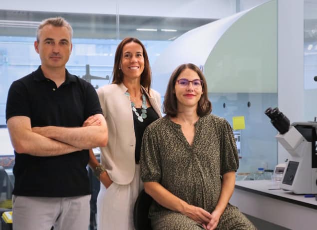
Samuel Ojosnegros, Anna Seriola and Amélie Godeau
Research team From left to right: Samuel Ojosnegros, Anna Seriola and Amélie Godeau at IBEC labs. (Courtesy: Institute for Bioengineering of Catalonia)

The researchers also monitored the traction forces that the embryos exerted on the collagen matrix, moving and reorganising it with a displacement that increased over time. They note that the displacement was not perfectly uniform and that the pulling varied over time and space, suggesting that this pulsatile behaviour may help the embryos to continuously sense the environment.

“We have observed that human embryos burrow into the uterus, exerting considerable force during the process,” explains study leader Samuel Ojosnegros in a press statement. “These forces are necessary because the embryos must be able to invade the uterine tissue, becoming completely integrated with it. It is a surprisingly invasive process. Although it is known that many women experience abdominal pain and slight bleeding during implantation, the process itself had never been observed before.”

For comparison, the researchers also examined the implantation of mouse blastocysts. In contrast to the complete integration seen for human blastocysts, mouse embryo outgrowth was limited to the matrix surface. In both platforms, initial attachment was followed by invasion and proliferation of trophoblast cells (the outer layer of the blastocyst). The embryo applied strong pulling forces to the fibrous matrix, remodelling the collagen and aligning the fibres around it during implantation. The displacement maps revealed a fluctuating pattern, as seen for the human embryos.

“By measuring the direct impact of the embryo on the matrix scaffold, we reveal the underlying mechanics of embryo implantation,” the researchers write. “We found that mouse and human embryos generated forces during implantation using a species-specific pattern.”

The team is now working to incorporate a theoretical framework to better understand the physical processes underlying implantation. “Our observations at earlier stages show that attachment is a limiting factor at the onset of human embryo implantation,” co-first author Amélie Godeau tells Physics World. “Our next step is to identify the key elements that enable a successful initial connection between the embryo and the matrix.”

The study is reported in Science Advances.

The post Researchers perform first real-time visualization of human embryo implantation appeared first on Physics World.

  •  

Squid use Bragg reflectors in their skin to change colour

Cephalopods such as squid and octopus can rapidly change the colour of their skin, but the way they do it has been something of a mystery – until now. Using a microscopy technique known as holotomography, scientists in the US discovered that the tuneable optical properties of squid skin stem from winding columns of platelets in certain cells. These columns have sinusoidal-wave refractive index profiles, and they function as Bragg reflectors, able to selectively transmit and reflect light at specific wavelengths.

“Our new result not only helps advance our understanding of structural colouration in cephalopods skin cells, it also provides new insights into how such gradient refractive index distributions can be leveraged to manipulate light in both biological and engineered systems,” says Alon Gorodetsky of the University of California, Irvine, who co-led this research study together with then-PhD student Georgii Bogdanov.

Stacked and winding columns of platelets

In their study, Gorodetsky, Bogdanov and colleagues including Roger Hanlon of the Marine Biological Laboratory (MBL) in Woods Hole, Massachusetts, examined the iridescent cells (iridophores) and cell clusters (splotches) responsible for producing colours in longfin inshore squids (Doryteuthis pealeii). To do this, they used holotomography, which creates three-dimensional images of individual cells and cell clusters by measuring subtle changes in a light beam as it passes through a sample of tissue. From this, they were able to map out changes in the sample’s refractive index across different structures.

The holotomography images revealed that the iridophores comprise stacked and winding columns of platelets made from a protein known as reflectin, which has a high refractive index, alternating with a low-refractive-index extracellular space. These Bragg-reflector-like structures are what allow tissue in the squid’s mantle to switch from nearly transparent to vibrantly coloured and back again.

Other natural Bragg reflectors

Squids aren’t the only animals that use Bragg reflectors for structural colouration, Gorodetsky notes. The scales of Morpho butterflies, for example, get their distinctive blue colouration from nanostructured Bragg gratings made from alternating high-refractive-index lamellae and low-refractive-index air gaps. Another example is the panther chameleon. The skin cells of this famously colour-changing reptile contain reconfigurable photonic lattices consisting of high-refractive-index nanocrystals within a low-refractive-index cytoplasm. These structures allow the animal to regulate its temperature as well as change its colour.

Yet despite these previous findings, and extensive research on cephalopod colouration, Gorodetsky says the question of how squid splotch iridophores can change from transparent to colourful , while maintaining their spectral purity, had not previously been studied in such depth. “In particular, the cells’ morphologies and refractive index distributions in three dimensions had not been previously resolved,” he explains. “Overcoming the existing knowledge gap required the development and application of combined experimental and computational approaches, including advanced imaging, refractive index mapping and optical modelling.”

Extending to infrared wavelengths

After using advanced computational modelling to capture the optical properties of the squid cells, the researchers, who report their work in Science, built on this result by designing artificial nanomaterials inspired by the natural structures they discovered. While the squid iridophores only change their visible appearance in response to neurophysiological stimuli, the researchers’ elastomeric composite materials (which contain both nanocolumnar metal oxide Bragg reflectors and nanostructured metal films) also change at infrared wavelengths.

Composite materials like the ones the UC Irvine-MBL team developed could have applications in adaptive camouflage or fabrics that adjust to hot and cold temperatures. They might also be used to improve multispectral displays, sensors, lasers, fibre optics and photovoltaics, all of which exploit multilayered Bragg reflectors with sinusoidal-wave refractive index profiles, says Gorodetsky.

The researchers now plan to further explore how gradient refractive index distributions contribute to light manipulation in other biological systems. “We also hope to refine our engineered multispectral composite materials to enhance their performance for specific practical applications, such as advanced camouflage and other wearable optical technologies,” Gorodetsky tells Physics World.

The post Squid use Bragg reflectors in their skin to change colour appeared first on Physics World.

  •  

Hyperdisorder appears in pigment patterns on squid skin

Researchers at the Okinawa Institute of Science and Technology (OIST) in Japan have identified the first known example of hyperdisorder occurring in a biological system. This phenomenon combines order at the microscopic scale with disorder at the macro level, and it is often present in systems studied in statistical physics. However, the researchers were surprised to observe it while monitoring the development of pigment cells in squid skin. As the hyperdisorder is directly linked to the squid’s growth, the researchers say the discovery could shed light on the physics of growing structures.

In inanimate objects, the emergence of disordered patterns is relatively well understood in physical terms. Living creatures are different, however, as they can display unexpected phenomena as they grow and develop.

To better understand how growth impacts the formation of patterns, a team led by Robert Ross, Simone Pigolotti and Sam Reiter at OIST studied how pigment cells known as chromatophores arrange themselves on the skin of squid as the animal grows and its skin expands. “These pigment cells are important because they play an essential role in camouflage and communication for these animals,” Reiter explains.

Highly unusual statistical patterns

The researchers took a series of 3D optical images of the squid over a period of three months. These observations revealed that the chromatophores behave very differently from other disordered structures. “The chromatophores appear at fixed positions in relation to one another, in a specific pattern,” Reiter explains.

It is this pattern that met the technical criteria for hyperdisorder, which is defined as occurring when the variation in the number of points within a particular measured space increases more rapidly than the volume of that space.

In the squid he and his colleagues studied, Ross explains that new chromatophores appear only at a minimum exclusion distance from pre-existing ones as the animal grows. “We found that this rule coupled with tissue growth leads to the highly unusual statistical patterns we observe,” he says. “Simply put, when you observe a tiny area in a system, it may appear quite ordered, but when viewed at larger scales, it becomes more disordered.”

To explain this finding, the researchers modelled squid development as static circle packing on a growing surface and showed how the hyperdisordered behaviour emerges. “The result is exciting because it highlights the importance of growth on physical properties,” Ross says.

A general feature of many biological structures?

The researchers note that other growing systems, such as the cells in chicken retinas, often display the exact opposite property, which is known as hyperuniformity. In these systems, there is long-range order and patterning despite randomness at a close scale, Ross explains. Such behaviour is thought to provide optimal retinal coverage properties for vision. “This is what we thought we would see in the squid, but what we actually observed was quite different and we have not yet seen any other instances of this packing behaviour in biology,” he says.

The mechanisms described in this work, which is detailed in Physical Review X, may be common in growing, dense natural systems, says Ross: “Indeed, this simple type of growth combined with distance-limited cell insertion might be a general feature of many biological structures.”

Spurred on by their findings, the researchers plan to continue working on a variety of theoretical and experimental systems related to the physics of growing structures. “These include both growing brains and pattern formation in fish,” says Ross.  “We hope these systems will provide further examples of the novel physics of growing systems,” he tells Physics World.

The post Hyperdisorder appears in pigment patterns on squid skin appeared first on Physics World.

  •