↩ Accueil

Vue lecture

Light-activated pacemaker is smaller than a grain of rice

The world’s smallest pacemaker to date is smaller than a single grain of rice, optically controlled and dissolves after it’s no longer needed. According to researchers involved in the work, the pacemaker could work in human hearts of all sizes that need temporary pacing, including those of newborn babies with congenital heart defects.

“Our major motivation was children,” says Igor Efimov, a professor of medicine and biomedical engineering, in a press release from Northwestern University. Efimov co-led the research with Northwestern bioelectronics pioneer John Rogers.

“About 1% of children are born with congenital heart defects – regardless of whether they live in a low-resource or high-resource country,” Efimov explains. “Now, we can place this tiny pacemaker on a child’s heart and stimulate it with a soft, gentle, wearable device. And no additional surgery is necessary to remove it.”

The current clinical standard-of-care involves sewing pacemaker electrodes directly onto a patient’s heart muscle during surgery. Wires from the electrodes protrude from the patient’s chest and connect to an external pacing box. Placing the pacemakers – and removing them later – does not come without risk. Complications include infection, dislodgment, torn or damaged tissues, bleeding and blood clots.

To minimize these risks, the researchers sought to develop a dissolvable pacemaker, which they introduced in Nature Biotechnology in 2021. By varying the composition and thickness of materials in the devices, Rogers’ lab can control how long the pacemaker functions before dissolving. The dissolvable device also eliminates the need for bulky batteries and wires.

“The heart requires a tiny amount of electrical stimulation,” says Rogers in the Northwestern release. “By minimizing the size, we dramatically simplify the implantation procedures, we reduce trauma and risk to the patient, and, with the dissolvable nature of the device, we eliminate any need for secondary surgical extraction procedures.”

Light-controlled pacing
Light-controlled pacing When the wearable device (left) detects an irregular heartbeat, it emits light to activate the pacemaker. (Courtesy: John A Rogers/Northwestern University)

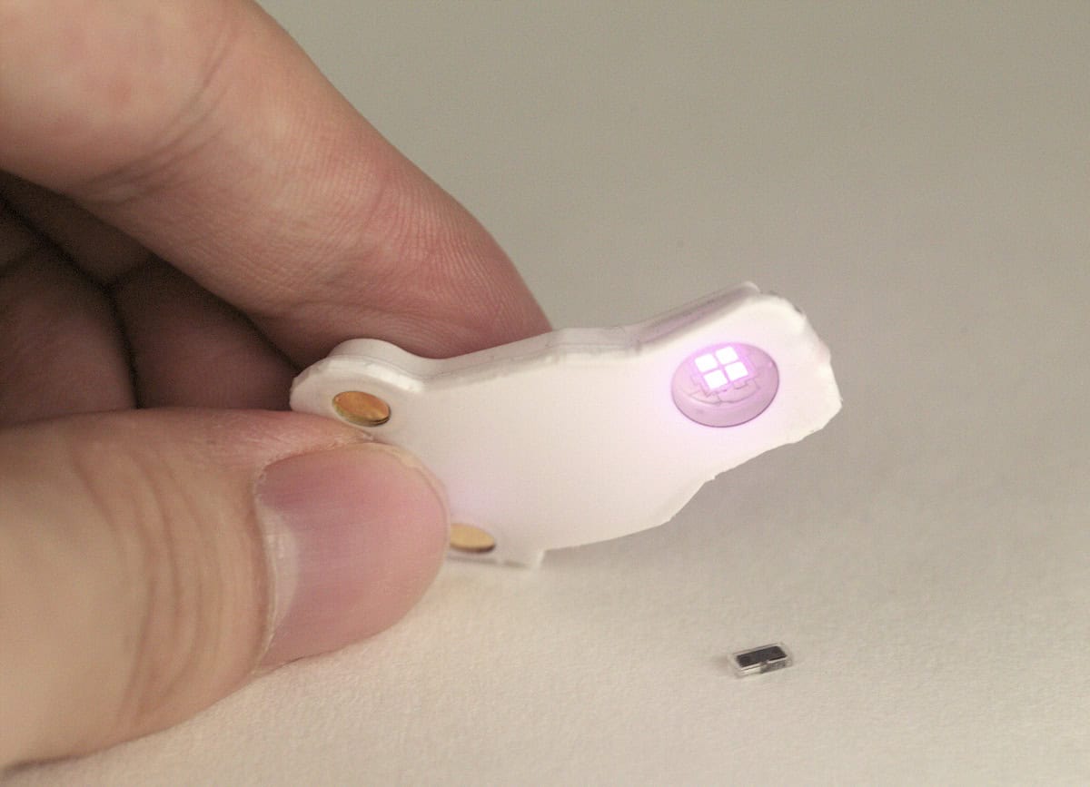
The latest iteration of the device – reported in Nature – advances the technology further. The pacemaker is paired with a small, soft, flexible, wireless device that is mounted onto the patient’s chest. The skin-interfaced device continuously captures electrocardiogram (ECG) data. When it detects an irregular heartbeat, it automatically shines a pulse of infrared light to activate the pacemaker and control the pacing.

“The new device is self-powered and optically controlled – totally different than our previous devices in those two essential aspects of engineering design,” says Rogers. “We moved away from wireless power transfer to enable operation, and we replaced RF wireless control strategies – both to eliminate the need for an antenna (the size-limiting component of the system) and to avoid the need for external RF power supply.”

Measurements demonstrated that the pacemaker – which is 1.8 mm wide, 3.5 mm long and 1 mm thick – delivers as much stimulation as a full-sized pacemaker. Initial studies in animals and in the human hearts of organ donors suggest that the device could work in human infants and adults. The devices are also versatile, the researchers say, and could be used across different regions of the heart or the body. They could also be integrated with other implantable devices for applications in nerve and bone healing, treating wounds and blocking pain.

The next steps for the research (supported by the Querrey Simpson Institute for Bioelectronics, the Leducq Foundation and the National Institutes of Health) include further engineering improvements to the device. “From the translational standpoint, we have put together a very early-stage startup company to work individually and/or in partnerships with larger companies to begin the process of designing the device for regulatory approval,” Rogers says.

The post Light-activated pacemaker is smaller than a grain of rice appeared first on Physics World.

  •  

Retinal stimulation reveals colour never before seen by the human eye

A new retinal stimulation technique called Oz enabled volunteers to see colours that lie beyond the natural range of human vision. Developed by researchers at UC Berkeley, Oz works by stimulating individual cone cells in the retina with targeted microdoses of laser light, while compensating for the eye’s motion.

Colour vision is enabled by cone cells in the retina. Most humans have three types of cone cells, known as L, M and S (long, medium and short), which respond to different wavelengths of visible light. During natural human vision, the spectral distribution of light reaching these cone cells determines the colours that we see.

Spectral sensitivity curves
Spectral sensitivity curves The response function of M cone cells overlaps completely with those of L and S cones. (Courtesy: Ben Rudiak-Gould)

Some colours, however, simply cannot be seen. The spectral sensitivity curves of the three cone types overlap – in particular, there is no wavelength of light that stimulates only the M cone cells without stimulating nearby L (and sometimes also S) cones as well.

The Oz approach, however, is fundamentally different. Rather than being based on spectral distribution, colour perception is controlled by shaping the spatial distribution of light on the retina.

Describing the technique in Science Advances, Ren Ng and colleagues showed that targeting individual cone cells with a 543 nm laser enabled subjects to see a range of colours in both images and videos. Intriguingly, stimulating only the M cone cells sent a colour signal to the brain that never occurs in natural vision.

The Oz laser system uses a technique called adaptive optics scanning light ophthalmoscopy (AOSLO) to simultaneously image and stimulate the retina with a raster scan of laser light. The device images the retina with infrared light to track eye motion in real time and targets pulses of visible laser light at individual cone cells, at a rate of 105 per second.

In a proof-of-principle experiment, the researchers tested a prototype Oz system on five volunteers. In a preparatory step, they used adaptive optics-based optical coherence tomography (AO-OCT) to classify the LMS spectral type of 1000 to 2000 cone cells in a region of each subject’s retina.

When exclusively targeting M cone cells in these retinal regions, subjects reported seeing a new blue–green colour of unprecedented saturation – which the researchers named “olo”. They could also clearly perceive Oz hues in image and video form, reliably detecting the orientation of a red line and the motion direction of a rotating red dot on olo backgrounds. In colour matching experiments, subjects could only match olo with the closest monochromatic light by desaturating it with white light – demonstrating that olo lies beyond the range of natural vision.

The team also performed control experiments in which the Oz microdoses were intentionally “jittered” by a few microns. With the target locations no longer delivered accurately, the subjects instead perceived the natural colour of the stimulating laser. In the image and video recognition experiments, jittering the microdose target locations reduced the task accuracy to guessing rate.

Ng and colleagues conclude that “Oz represents a new class of experimental platform for vision science and neuroscience [that] will enable diverse new experiments”. They also suggest that the technique could one day help to elicit full colour vision in people with colour blindness.

The post Retinal stimulation reveals colour never before seen by the human eye appeared first on Physics World.

  •  

Hydrogels rapidly switch from soft to hard to create smart medical bandage

Hard/soft transition of a hydrogel/NAAC composite
Reversible switching Schematic illustrating the hard/soft transition of the hydrogel/NAAC composite. (Courtesy: CC BY 4.0/Int. J. Extrem. Manuf. 10.1088/2631-7990/adbd97)

Complex hydrogel structures created using 3D printing are increasingly employed in fields including flexible electronics, soft robotics and regenerative medicine. Currently, however, such hydrogels are often soft and fragile, limiting their practical utility. Researchers at Zhejiang University in China have now fabricated 3D-printed hydrogels that can be easily, and repeatably, switched between soft and hard states, enabling novel applications such as smart medical bandages or information encryption.

“Our primary motivation was to overcome the inherent limitations of 3D-printed hydrogels, particularly their soft, weak and fragile mechanical properties, to broaden their application potential,” says co-senior author Yong He.

The research team created the hard/soft switchable composite by infusing supersaturated salt solution (sodium acetate, NAAC) into 3D-printed polyacrylamide (PAAM)-based hydrogel structures. The hardness switching is enabled by the liquid/solid transition of the salt solution within the hydrogel.

Initially, the salt molecules are arranged randomly within the hydrogel and the PAAM/NAAC composite is soft and flexible. The energy barrier separating the soft and hard states prevents spontaneous crystallization, but can be overcome by artificially seeding a crystal nucleus (via exposure to a salt crystal or contact with a sharp object). This seed promotes a phase transition to a hard state, with numerous rigid, rod-like nanoscale crystals forming within the hydrogel matrix.

Superior mechanical parameters

The researchers created a series of PAAM/NAAC structures, using projection-based 3D printing to print hydrogel shapes and then soaking them in NAAC solution. Upon seeding, the structures rapidly transformed from transparent to opaque as the crystallization spread through the sample at speeds of up to 4.5 mm/s.

The crystallization dramatically changed the material’s mechanical performance. For example, a soft cylinder of PAAM/1.5NAAC (containing 150 wt% salt) could be easily compressed by hand, returning to its original shape after release. After crystallization, four 9x9x12 mm cylinders could support an adult’s weight without deforming.

For this composite, just 1 min of crystallization dramatically increased the compression Young’s modulus compared with the soft state. And after 24 h, the Young’s modulus grew from 110 kPa to 871.88 MPa. Importantly, the hydrogel could be easily returned to its soft state by heating and then cooling, a process that could be repeated many times.

The team also performed Shore hardness testing on various composites, observing that hardness values increased with increasing NAAC concentration. In PAAM/1.7NAAC composites (170 wt% salt), the Shore D value reached 86.5, comparable to that of hard plastic materials.

The hydrogel’s crosslinking density also impacted its mechanical performance. For PAAM/1.5NAAC composites, increasing the mass percentage of polymer crosslinker from 0.02 to 0.16 wt% increased the compression Young’s modulus to 1.2 GPa and the compression strength to 81.7 MPa. The team note that these parameters far exceed those of any existing 3D-printed hydrogels.

Smart plaster cast

He and colleagues demonstrated how the hard/soft switching and robust mechanical properties of PAAM/NAAC can create medical fixation devices, such as a smart plaster cast. The idea here is that the soft hydrogel can be moulded around the injured bone, and then rapidly frozen in shape by crystallization to support the injury and promote healing.

The researchers tested the smart plaster cast on an injured forearm. After applying a layer of soft cotton padding, they carefully wrapped around layers of the smart plaster bandage (packed within a polyethylene film to prevent accidental seeding). The flexible hydrogel could be conformed to the curved surface of limbs and then induced to crystallize.

Smart plaster cast
Medical fixation device Application of the PAAM/NAAC composite to create a smart plaster cast. (Courtesy: CC BY 4.0/Int. J. Extrem. Manuf. 10.1088/2631-7990/adbd97)

After just 10 min of crystallization, the smart plaster cast reached a yield strength of 8.7 MPa, rapidly providing support for the injured arm. In comparison, a traditional plaster cast (as currently used to treat bone fractures) took about 24 h to fully harden, reaching a maximum yield strength of 3.9 MPa

To determine the safety of the exothermic crystallization process, the team monitored temperature changes in the plaster cast nearest to the skin. The temperature peaked at 41.5 °C after 25 min of crystallization, below the ISO-recommended maximum safe temperature of 50 °C.

The researchers suggest that the ease of use, portability and fast response of the smart plaster cast could provide a simple and effective solution for emergency and first aid situations. Another benefit is that, in contrast to traditional plaster casts that obstruct X-rays and hinder imaging, X-rays easily penetrate through the smart plaster cast to enable high-quality imaging during the healing process.

While the composites exhibit high strength and Young’s modulus, they are not as tough as ideally desired. “For example, the elongation at break was less than 10% in tensile testing for the PAAM/1.5NAAC and PAAM/1.7NAAC samples, highlighting the challenge of balancing toughness with strength and modulus,” He tells Physics World. “Therefore, our current research focuses on enhancing the toughness of these composite materials without compromising their modulus, with the goal of developing strong, tough and mechanically switchable materials.”

The hydrogel is described in the International Journal of Extreme Manufacturing.

The post Hydrogels rapidly switch from soft to hard to create smart medical bandage appeared first on Physics World.

  •  

Zwitterions make medical implants safer for patients

A new technique could reduce the risk of blood clots associated with medical implants, making them safer for patients. The technique, which was developed by researchers at the University of Sydney, Australia, involves coating the implants with highly hydrophilic molecules known as zwitterions, thereby inhibiting the build-up of clot-triggering proteins.

Proteins in blood can stick to the surfaces of medical implants such as heart valves and vascular stents. When this happens, it produces a cascade effect in which multiple mechanisms lead to the formation of extensive clots and fibrous networks. These clots and networks can impair the function of implanted medical devices so much that invasive surgery may be required to remove or replace the implant.

To prevent this from happening, the surfaces of implants are often treated with polymeric coatings that resist biofouling. Hydrophilic polymeric coatings such as polyethylene glycol are especially useful, as their water-loving nature allows a thin layer of water to form between them and the surface of the implants, held in place via hydrogen and/or electrostatic bonds. This water layer forms a barrier that prevents proteins from sticking, or adsorbing, to the implant.

An extra layer of zwitterions

Recently, researchers discovered that polymers coated with an extra layer of small molecules called zwitterions provided even more protection against protein adsorption. “Zwitter” means “hybrid” in German; hence, zwitterions are molecules that carry both positive and negative charge, making them neutrally charged overall. These molecules are also very hydrophilic and easily form tight bonds with water molecules. The resulting layer of water has a structure that is similar to that of bulk water, which is energetically stable.

A further attraction of zwitterionic coatings for medical implants is that zwitterions are naturally present in our bodies. In fact, they make up the hydrophilic phospholipid heads of mammalian cell membranes, which play a vital role in regulating interactions between biological cells and the extracellular environment.

Plasma functionalization

In the new work, researchers led by Sina Naficy grafted nanometre-thick zwitterionic coatings onto the surfaces of implant materials using a technique called plasma functionalization. They found that the resulting structures reduce the amount of fibrinogen proteins that adsorb onto the implants by roughly nine-fold and decrease blood clot formation (thrombosis) by almost 75%.

Naficy and colleagues achieved their results by optimizing the density, coverage and thickness of the coating. This was critical for realizing the full potential of these materials, they say, because a coating that is not fully optimized would not reduce clotting.

Naficy tells Physics World that the team’s main goal is to enhance the surface properties of medical devices. “These devices when implanted are in contact with blood and can readily cause thrombosis or infection if the surface initiates certain biological cascade reactions,” he explains. “Most such reactions begin when specific proteins adsorb on the surface and activate the next stage of cascade. Optimizing surface properties with the aid of zwitterions can control / inhibit protein adsorption, hence reducing the severity of adverse body reactions.”

The researchers say they will now be evaluating the long-term stability of the zwitterion-polymer coatings and trying to scale up their grafting process. They report their work in Communications Materials and Cell Biomaterials.

The post Zwitterions make medical implants safer for patients appeared first on Physics World.

  •  

Novel zinc alloys could make bone screws biodegradable

Orthopaedic implants that bear loads while bones heal, then disappear once they’re no longer needed, could become a reality thanks to a new technique for enhancing the mechanical properties of zinc alloys. Developed by researchers at Monash University in Australia, the technique involves controlling the orientation and size of microscopic grains in these strong yet biodegradable materials.

Implants such as plates and screws provide temporary support for fractured bones until they knit together again. Today, these implants are mainly made from sturdy materials such as stainless steel or titanium that remain in the body permanently. Such materials can, however, cause discomfort and bone loss, and subsequent injuries to the same area risk additional damage if the permanent implants warp or twist.

To address these problems, scientists have developed biodegradable alternatives that dissolve once the bone has healed. These alternatives include screws made from magnesium-based materials such as MgYREZr (trade name MAGNEZIX), MgYZnMn (NOVAMag) and MgCaZn (RESOMET). However, these materials have compressive yield strengths of just 50 to 260 MPa, which is too low to support bones that need to bear a patient’s weight. They also produce hydrogen gas as they degrade, possibly affecting how biological tissues regenerate.

Zinc alloys do not suffer from the hydrogen gas problem. They are biocompatible, dissolving slowly and safely in the body. There is even evidence that Zn2+ ions can help the body heal by stimulating bone formation. But again, their mechanical strength is low: at less than 30 MPa, they are even worse than magnesium in this respect.

Making zinc alloys strong enough for load-bearing orthopaedic implants is not easy. Mechanical strategies such as hot-extruding binary alloys have not helped much. And methods that focus on reducing the materials’ grain size (to hamper effects like dislocation slip) have run up against a discouraging problem: at body temperature (37 °C), ultrafine-grained Zn alloys become mechanically weaker as their so-called “creep resistance” decreases.

Grain size goes bigger

In the new work, a team led by materials scientist and engineer Jian-Feng Nei tried a different approach. By increasing grain size in Zn alloys rather than decreasing it, the Monash team was able to balance the alloys’ strength and creep resistance – something they say could offer a route to stronger zinc alloys for biodegradable implants.

In compression tests of extruded Zn–0.2 wt% Mg alloy samples with grain sizes of 11 μm, 29 μm and 47 μm, the team measured stress-strain curves that show a markedly higher yield strength for coarse-grained samples than for fine-grained ones. What is more, the compressive yield strengths of these coarser-grained zinc alloys are notably higher than those of MAGNEZIX, NOVAMag and RESOMET biodegradable magnesium alloys. At the upper end, they even rival those of high-strength medical-grade stainless steels.

The researchers attribute this increased compressive yield to a phenomenon called the inverse Hall–Petch effect. This effect comes about because larger grains favour metallurgical effects such as intra-granular pyramidal slip as well as a variation of a well-known metal phenomenon called twinning, in which a specific kind of defect forms when part of the material’s crystal structure flips its orientation. Larger grains also make the alloys more flexible, allowing them to better adapt to surrounding biological tissues. This is the opposite of what happens with smaller grains, which facilitate inter-granular grain boundary sliding and make alloys more rigid.

The new work, which is detailed in Nature, could aid the development of advanced biodegradable implants for orthopaedics, cardiovascular applications and other devices, says Nei. “With improved biocompatibility, these implants could be safer and do away with the need for removal surgeries, lowering patient risk and healthcare costs,” he tells Physics World. “What is more, new alloys and processing techniques could allow for more personalized treatments by tailoring materials to specific medical needs, ultimately improving patient outcomes.”

The Monash team now aims to improve the composition of the alloys and achieve more control over how they degrade. “Further studies on animals and then clinical trials will test their strength, safety and compatibility with the body,” says Nei. “After that, regulatory approvals will ensure that the biodegradable metals meet medical standards for orthopaedic implants.”

The team is also setting up a start-up company with the goal of developing and commercializing the materials, he adds.

The post Novel zinc alloys could make bone screws biodegradable appeared first on Physics World.

  •  

Cell sorting device could detect circulating tumour cells

Acousto-microfluidic chip
Cell separation Illustration of the fabricated optimal acousto-microfluidic chip. (Courtesy: Afshin Kouhkord and Naserifar Naser)

Analysing circulating tumour cells (CTCs) in the blood could help scientists detect cancer in the body. But separating CTCs from blood is a difficult, laborious process and requires large sample volumes.

Researchers at the K N Toosi University of Technology (KNTU) in Tehran, Iran believe that ultrasonic waves could separate CTCs from red blood cells accurately, in an energy efficient way and in real time. They publish their study in the journal Physics of Fluids.

“In a broader sense, we asked: ‘How can we design a microfluidic, lab-on-a-chip device powered by SAWs [standing acoustic waves] that remains simple enough for medical experts to use easily, while still delivering precise and efficient cell separation?’,” says senior author Naser Naserifar, an assistant professor in mechanical engineering at KNTU. “We became interested in acoustofluidics because it offers strong, biocompatible forces that effectively handle cells with minimal damage.”

Acoustic waves can deliver enough force to move cells over small distances without damaging them. The researchers used dual pressure acoustic fields at critical positions in a microchannel to separate CTCs from other cells. The CTCs are gathered at an outlet for further analyses, cultures and laboratory procedures.

In the process of designing the chip, the researchers integrated computational modelling, experimental analysis and artificial intelligence (AI) algorithms to analyse acoustofluidic phenomena and generate datasets that predict CTC migration in the body.

“We introduced an acoustofluidic microchannel with two optimized acoustic zones, enabling fast, accurate separation of CTCs from RBCs [red blood cells],” explains Afshin Kouhkord, who performed the work while a master’s student in the Advance Research in Micro And Nano Systems Lab at KNTU. “Despite the added complexity under the hood, the resulting chip is designed for simple operation in a clinical environment.”

So far, the researchers have evaluated the device with numerical simulations and tested it using a physical prototype. Simulations modelled fluid flow, acoustic pressure fields and particle trajectories. The physical prototype was made of lithium niobate, with polystyrene microspheres used as surrogates for red blood cells and CTCs. Results from the prototype agreed with numerical simulations to within 3.5%.

“This innovative approach in laboratory-on-chip technology paves the way for personalized medicine, real-time molecular analysis and point-of-care diagnostics,” Kouhkord and Naserifar write.

The researchers are now refining their design, aiming for a portable device that could be operated with a small battery pack in resource-limited and remote environments.

The post Cell sorting device could detect circulating tumour cells appeared first on Physics World.

  •  

Nanosensor predicts risk of complications in early pregnancy

Researchers in Australia have developed a nanosensor that can detect the onset of gestational diabetes with 95% accuracy. Demonstrated by a team led by Carlos Salomon at the University of Queensland, the superparamagnetic “nanoflower” sensor could enable doctors to detect a variety of complications in the early stages of pregnancy.

Many complications in pregnancy can have profound and lasting effects on both the mother and the developing foetus. Today, these conditions are detected using methods such as blood tests, ultrasound screening and blood pressure monitoring. In many cases, however, their sensitivity is severely limited in the earliest stages of pregnancy.

“Currently, most pregnancy complications cannot be identified until the second or third trimester, which means it can sometimes be too late for effective intervention,” Salomon explains.

To tackle this challenge, Salomon and his colleagues are investigating the use of specially engineered nanoparticles to isolate and detect biomarkers in the blood associated with complications in early pregnancy. Specifically, they aim to detect the protein molecules carried by extracellular vesicles (EVs) – tiny, membrane-bound particles released by the placenta, which play a crucial role in cell signalling.

In their previous research, the team pioneered the development of superparamagnetic nanostructures that selectively bind to specific EV biomarkers. Superparamagnetism occurs specifically in small, ferromagnetic nanoparticles, causing their magnetization to randomly flip direction under the influence of temperature. When proteins are bound to the surfaces of these nanostructures, their magnetic responses are altered detectably, providing the team with a reliable EV sensor.

“This technology has been developed using nanomaterials to detect biomarkers at low concentrations,” explains co-author Mostafa Masud. “This is what makes our technology more sensitive than current testing methods, and why it can pick up potential pregnancy complications much earlier.”

Previous versions of the sensor used porous nanocubes that efficiently captured EVs carrying a key placental protein named PLAP. By detecting unusual levels of PLAP in the blood of pregnant women, this approach enabled the researchers to detect complications far more easily than with existing techniques. However, the method generally required detection times lasting several hours, making it unsuitable for on-site screening.

In their latest study, reported in Science Advances, Salomon’s team started with a deeper analysis of the EV proteins carried by these blood samples. Through advanced computer modelling, they discovered that complications can be linked to changes in the relative abundance of PLAP and another placental protein, CD9.

Based on these findings, they developed a new superparamagnetic nanosensor capable of detecting both biomarkers simultaneously. Their design features flower-shaped nanostructures made of nickel ferrite, which were embedded into specialized testing strips to boost their sensitivity even further.

Using this sensor, the researchers collected blood samples from 201 pregnant women at 11 to 13 weeks’ gestation. “We detected possible complications, such as preterm birth, gestational diabetes and preeclampsia, which is high blood pressure during pregnancy,” Salomon describes. For gestational diabetes, the sensor demonstrated 95% sensitivity in identifying at-risk cases, and 100% specificity in ruling out healthy cases.

Based on these results, the researchers are hopeful that further refinements to their nanoflower sensor could lead to a new generation of EV protein detectors, enabling the early diagnosis of a wide range of pregnancy complications.

“With this technology, pregnant women will be able to seek medical intervention much earlier,” Salomon says. “This has the potential to revolutionize risk assessment and improve clinical decision-making in obstetric care.”

The post Nanosensor predicts risk of complications in early pregnancy appeared first on Physics World.

  •