↩ Accueil

Vue lecture

Hydrogels rapidly switch from soft to hard to create smart medical bandage

Hard/soft transition of a hydrogel/NAAC composite
Reversible switching Schematic illustrating the hard/soft transition of the hydrogel/NAAC composite. (Courtesy: CC BY 4.0/Int. J. Extrem. Manuf. 10.1088/2631-7990/adbd97)

Complex hydrogel structures created using 3D printing are increasingly employed in fields including flexible electronics, soft robotics and regenerative medicine. Currently, however, such hydrogels are often soft and fragile, limiting their practical utility. Researchers at Zhejiang University in China have now fabricated 3D-printed hydrogels that can be easily, and repeatably, switched between soft and hard states, enabling novel applications such as smart medical bandages or information encryption.

“Our primary motivation was to overcome the inherent limitations of 3D-printed hydrogels, particularly their soft, weak and fragile mechanical properties, to broaden their application potential,” says co-senior author Yong He.

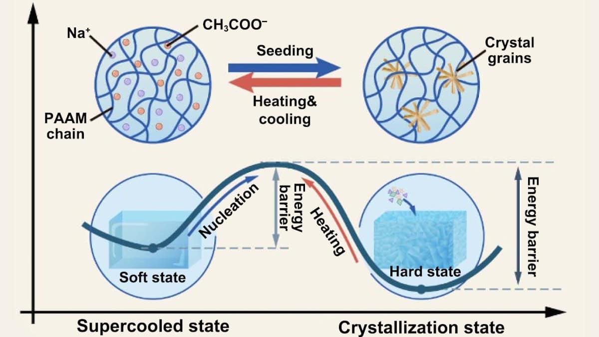
The research team created the hard/soft switchable composite by infusing supersaturated salt solution (sodium acetate, NAAC) into 3D-printed polyacrylamide (PAAM)-based hydrogel structures. The hardness switching is enabled by the liquid/solid transition of the salt solution within the hydrogel.

Initially, the salt molecules are arranged randomly within the hydrogel and the PAAM/NAAC composite is soft and flexible. The energy barrier separating the soft and hard states prevents spontaneous crystallization, but can be overcome by artificially seeding a crystal nucleus (via exposure to a salt crystal or contact with a sharp object). This seed promotes a phase transition to a hard state, with numerous rigid, rod-like nanoscale crystals forming within the hydrogel matrix.

Superior mechanical parameters

The researchers created a series of PAAM/NAAC structures, using projection-based 3D printing to print hydrogel shapes and then soaking them in NAAC solution. Upon seeding, the structures rapidly transformed from transparent to opaque as the crystallization spread through the sample at speeds of up to 4.5 mm/s.

The crystallization dramatically changed the material’s mechanical performance. For example, a soft cylinder of PAAM/1.5NAAC (containing 150 wt% salt) could be easily compressed by hand, returning to its original shape after release. After crystallization, four 9x9x12 mm cylinders could support an adult’s weight without deforming.

For this composite, just 1 min of crystallization dramatically increased the compression Young’s modulus compared with the soft state. And after 24 h, the Young’s modulus grew from 110 kPa to 871.88 MPa. Importantly, the hydrogel could be easily returned to its soft state by heating and then cooling, a process that could be repeated many times.

The team also performed Shore hardness testing on various composites, observing that hardness values increased with increasing NAAC concentration. In PAAM/1.7NAAC composites (170 wt% salt), the Shore D value reached 86.5, comparable to that of hard plastic materials.

The hydrogel’s crosslinking density also impacted its mechanical performance. For PAAM/1.5NAAC composites, increasing the mass percentage of polymer crosslinker from 0.02 to 0.16 wt% increased the compression Young’s modulus to 1.2 GPa and the compression strength to 81.7 MPa. The team note that these parameters far exceed those of any existing 3D-printed hydrogels.

Smart plaster cast

He and colleagues demonstrated how the hard/soft switching and robust mechanical properties of PAAM/NAAC can create medical fixation devices, such as a smart plaster cast. The idea here is that the soft hydrogel can be moulded around the injured bone, and then rapidly frozen in shape by crystallization to support the injury and promote healing.

The researchers tested the smart plaster cast on an injured forearm. After applying a layer of soft cotton padding, they carefully wrapped around layers of the smart plaster bandage (packed within a polyethylene film to prevent accidental seeding). The flexible hydrogel could be conformed to the curved surface of limbs and then induced to crystallize.

Smart plaster cast
Medical fixation device Application of the PAAM/NAAC composite to create a smart plaster cast. (Courtesy: CC BY 4.0/Int. J. Extrem. Manuf. 10.1088/2631-7990/adbd97)

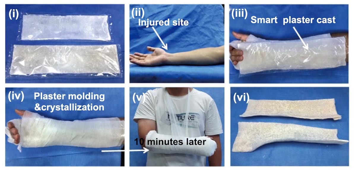
After just 10 min of crystallization, the smart plaster cast reached a yield strength of 8.7 MPa, rapidly providing support for the injured arm. In comparison, a traditional plaster cast (as currently used to treat bone fractures) took about 24 h to fully harden, reaching a maximum yield strength of 3.9 MPa

To determine the safety of the exothermic crystallization process, the team monitored temperature changes in the plaster cast nearest to the skin. The temperature peaked at 41.5 °C after 25 min of crystallization, below the ISO-recommended maximum safe temperature of 50 °C.

The researchers suggest that the ease of use, portability and fast response of the smart plaster cast could provide a simple and effective solution for emergency and first aid situations. Another benefit is that, in contrast to traditional plaster casts that obstruct X-rays and hinder imaging, X-rays easily penetrate through the smart plaster cast to enable high-quality imaging during the healing process.

While the composites exhibit high strength and Young’s modulus, they are not as tough as ideally desired. “For example, the elongation at break was less than 10% in tensile testing for the PAAM/1.5NAAC and PAAM/1.7NAAC samples, highlighting the challenge of balancing toughness with strength and modulus,” He tells Physics World. “Therefore, our current research focuses on enhancing the toughness of these composite materials without compromising their modulus, with the goal of developing strong, tough and mechanically switchable materials.”

The hydrogel is described in the International Journal of Extreme Manufacturing.

The post Hydrogels rapidly switch from soft to hard to create smart medical bandage appeared first on Physics World.

  •  

Simultaneous two- and three-photon imaging enables rapid cancer diagnosis

Multiphoton microscopy is a nonlinear optical imaging technique that enables label-free, damage-free biological imaging. Performed using femtosecond laser pulses to generate two- and three-photon processes, multiphoton imaging techniques could prove invaluable for rapid cancer diagnosis or personalized medicine.

Imaging biological samples with traditional confocal microscopy requires sample slicing and staining to create contrast in the tissue. The nonlinear mechanisms generated by femtosecond laser pulses, however, eliminate the need for labelling or sample preparation, revealing molecular and structural details within tissue and cells while leaving the sample intact.

Looking to bring these benefits to cancer diagnostics, Netherlands-based start-up Flash Pathology is developing a compact, portable multiphoton microscope that creates pathology-quality images in real time, without the need for sample fixation or staining.

Fast and accurate cancer diagnosis with higher harmonic imaging

The inspiration for Flash Pathology came from Marloes Groot of Vrije Universiteit (VU) Amsterdam. While studying multiphoton microscopy of brain tumours, Groot recognized the need for a portable microscope for clinical settings. “This is a really powerful technique,” she says. “I was working on my large laboratory setup and I thought if we start a company, we could transform this into a mobile device.”

Groot teamed up with Frank van Mourik, now Flash Pathology’s CTO, to shrink the imaging device into a compact 60 x 80 x 115 cm system. “Frank made a device that can be transported, in a truck, wheeled through corridors, and still when you plug it in, it’s on and it produces images – for a nonlinear microscope, this is pretty special,” she explains.

Van Mourik has now built several multiphoton microscopes for Flash Pathology, with one of the main achievements the ability to measure samples with extremely low power levels. “When I started in my lab, I used 200 mW of power, but we’ve been able to reduce that to 5 mW,” Groot notes. “We have performed extensive studies to show that our imaging does not affect the tissue.”

Flash Pathology’s multiphoton microscope is designed to provide rapid on-site histologic feedback on excised tissue, such as diagnostic biopsies or tissue from surgical resections. One key application is lung cancer diagnosis, where there is a clinical need for rapid intraoperative feedback on biopsies. The standard histopathological analysis requires extensive sample preparation and can take several days to provide results.

Multiphoton microscopy of adipose tissue, cartilage and lymphatic tissue
Rapid tissue analysis Multiphoton microscopy images of (left to right) adipose tissue, cartilage and lymphoid tissue (each image is 400 x 400 µm). The adipose tissue image shows large adipocytes (fat cells); the cartilage image shows a hyaline (glass-like) background with chondrocytes (cells); the image of lymphoid tissue shows many small lymphocytes (a type of immune cell). (Courtesy: VU Amsterdam)

“With lung biopsies, it’s challenging to obtain good diagnostic material,” explains Sylvia Spies, a PhD student at VU Amsterdam. “The lesions can be quite small and it can be difficult to get to the right position and take a good sample, so they use several techniques (fluoroscopy/CT or ultrasound) to find the right position and take multiple biopsies from the lesion. Despite these techniques, the diagnostic yield is still around 70%, so 30% of cases still don’t get a diagnosis and patients might have to come back for a repeat biopsy procedure.”

Multiphoton imaging, on the other hand, can rapidly visualize unprocessed tissue samples, enabling diagnosis in situ. A recent study using Flash Pathology’s microscope to analyse lung biopsies demonstrated that it could image a biopsy sample and provide feedback just 6 min after excision, with an accuracy of 87% – enabling immediate decisions as to whether a further biopsy is required.

“Also, many clinical fields are now focusing on a one-stop-shop with diagnosis and treatment in one procedure,” adds Spies. “Here, you really need a technique that can rapidly determine whether a lesion is benign or malignant.”

The microscope’s impressive diagnostic performance is partly due to its ability to generate four nonlinear signals simultaneously using a single ultrafast femtosecond laser: second- and third-harmonic generation plus two- and three-photon fluorescence. The system then uses filters to spectrally separate these signals, which provide complementary diagnostic information. Second-harmonic generation, for example, is sensitive to non-centrosymmetric structures such as collagen, while third-harmonic generation only occurs at interfaces with differing refractive indices, such as cell membranes or boundaries between the nucleus and cytoplasm.

“What I like about this technique is that you can see similar features as in conventional histology,” says Spies. “You can see structures such as collagen fibres, elastin fibres and cellular patterns, but also cellular details such as the cytoplasm, the nucleus (and its size), nucleoli and cilia. All these tiny details are the same features that the pathologists look at in conventional histology.”

Applying femtosecond lasers for 3D-in-depth visualization

The femtosecond laser plays a key role in enabling multiphoton microscopy. To excite two- and three-photon processes, you need to have two or more photons in the same place at exactly the same time. And the likelihood of this happening increases rapidly when using ultrashort laser pulses.

“The shorter the pulses are in the time domain, the higher the probability that you have an overlap of two pulses in a focal point,” explains Oliver Prochnow, CEO of VALO Innovations, a part of HÜBNER Photonics. “Therefore you need to have a very high-intensity, extremely short laser pulse. The shorter the better.”

The VALO Femtosecond Series of ultrafast fibre lasers can deliver pulses as short as 30 fs, which is achieved by exploiting nonlinear mechanisms to broaden the spectral bandwidth to more than 100 nm. As the optical spectrum and pulse duration are inherently related by Fourier transformation, a broadband spectrum will result in a very short pulse. And the shorter the pulse, at the same average power, the higher its peak power – and the higher the probability of producing multiphoton processes.

VALO Femtosecond Series laser parameters
Laser parameters Left: typical temporal pulse profile highlighting the sub 50 fs pulse duration with very low pulse pedestal; the inset shows the typical beam profile. Right: typical optical spectrum of HÜBNER Photonics’ VALO Femtosecond Series lasers. (Courtesy: HÜBNER Photonics)

“If you decrease the pulse duration by a factor of five, this gives roughly a five times higher signal from two-photon absorption,” says Prochnow. “In contrast, a three-photon process scales with the third power of the intensity and with the inverse of the pulse duration squared. So you have a roughly 25 times higher signal, if you decrease the pulse duration by a factor of five at the same average power.” Crucially, the shorter pulses deliver this high peak power while maintaining a low average power, reducing sample heating and minimizing photobleaching.

The broadband optical spectrum is particularly important for enabling practical three-photon microscopy. The challenge here is that traditional ytterbium-based lasers with a wavelength of around 1030 nm produce a three-photon signal in the UV range, which is too short to be transmitted through standard optics.

Third-harmonic generation spectra
Broadband spectrum Fundamental and third-harmonic generation (THG) spectra of a 30 fs broadband fibre laser (red) compared with standard 150 fs lasers. The solid black line shows the typical transmission characteristics of a standard microscopy objective. Only a THG spectrum generated from wavelengths of above 1080 nm will be transmitted. (Courtesy: HÜBNER Photonics)

The VALO Femtosecond Series overcomes this problem by having a broadband spectrum that extends up to 1140 nm. Frequency tripling then generates a signal with a long enough wavelength to pass through a standard microscope objective, enabling the VALO lasers to excite both two-photon and three-photon processes. “Our lasers provide the opportunity to perform simultaneous three-photon microscopy and two-photon microscopy using a simple fibre laser solution,” says Prochnow.

The lasers include an integrated dispersion pre-compensation unit to compensate for the dispersion of a microscope objective and provide the shortest pulses at the sample. Additionally, the lasers do not require water cooling, making them easy to use or integrate.

Towards future clinical applications

Flash Pathology is currently testing its microscope in several hospitals in the Netherlands, including Amsterdam UMC, as well as the Princess Maxima Center for paediatric oncology. “Sylvia performed a study in their pathology department and for a year measured all kinds of tissue samples that came through,” says Groot. “We also recently installed a device at the Queen Elizabeth Hospital in Glasgow, for a study on mesothelioma.”

With prototypes now available for research use, the company also plans to develop a fully certified multiphoton microscopy system. “Our ultimate goal is to sell a certified medical diagnostic device that will take a biopsy and produce images, but also contain artificial intelligence to help to interpret the images and give diagnostic conclusions about the nature of the illness,” says van Mourik.

Once fully realised in the clinic, the multiphoton microscopy system will provide an invaluable tool for rapid, in situ tissue analysis during bronchoscopy procedures or other operations. The unique combination of four nonlinear imaging modalities, made possible with a single compact femtosecond laser, delivers complementary diagnostic information. “This will be the big gain, to be able to provide a diagnosis bedside during a procedure,” van Mourik concludes.


The post Simultaneous two- and three-photon imaging enables rapid cancer diagnosis appeared first on Physics World.

  •  

Radioactive gold nanoparticles could track drug distribution in the body

Schematic showing activation imaging
Neutron-activated gold Novel activation imaging technique enables real-time visualization of gold nanoparticles in the body without the use of external tracers. (Courtesy: Nanase Koshikawa from Waseda University)

Gold nanoparticles are promising vehicles for targeted delivery of cancer drugs, offering biocompatibility plus a tendency to accumulate in tumours. To fully exploit their potential, it’s essential to be able to track the movement of these nanoparticles in the body. To date, however, methods for directly visualizing their pharmacokinetics have not yet been established. Aiming to address this shortfall, researchers in Japan are using neutron-activated gold radioisotopes to image nanoparticle distribution in vivo.

The team, headed up by Nanase Koshikawa and Jun Kataoka from Waseda University, are investigating the use of radioactive gold nanoparticles based on 198Au, which they create by irradiating stable gold (197Au) with low-energy neutrons. The radioisotope 198Au has a half-life of 2.7 days and emits 412 keV gamma rays, enabling a technique known as activation imaging.

“Our motivation was to visualize gold nanoparticles without labelling them with tracers,” explains Koshikawa. “Radioactivation allows gold nanoparticles themselves to become detectable from outside the body. We used neutron activation because it does not change the atomic number, ensuring the chemical properties of gold nanoparticles remain unchanged.”

In vivo studies

The researchers – also from Osaka University and Kyoto University – synthesized 198Au-based nanoparticles and injected them into tumours in four mice. They used a hybrid Compton camera (HCC) to detect the emitted 412 keV gamma rays and determine the in vivo nanoparticle distribution, on the day of injection and three and five days later.

The HCC, which incorporates two pixelated scintillators, a scatterer with a central pinhole, and an absorber, can detect radiation with energies from tens of keV to nearly 1 MeV. For X-rays and low-energy gamma rays, the scatterer enables pinhole-mode imaging. For gamma rays over 200 keV, the device functions as a Compton camera.

The researchers reconstructed the 412 keV gamma signals into images, using an energy window of 412±30 keV. With the HCC located 5 cm from the animals’ abdomens, the spatial resolution was 7.9 mm, roughly comparable to the tumour size on the day of injection (7.7 x 11 mm).

In vivo nanoparticle distribution
In vivo distribution Images of 198Au nanoparticles in the bodies of two mice obtained with the HCC on the day of administration. (Courtesy: CC BY 4.0/Appl. Phys. Lett. 10.1063/5.0251048)

Overlaying the images onto photographs of the mice revealed that the nanoparticles accumulated in both the tumour and liver. In mice 1 and 2, high pixel values were observed primarily in the tumour, while mice 3 and 4 also had high pixel values in the liver region.

After imaging, the mice were euthanized and the team used a gamma counter to measure the radioactivity of each organ. The measured activity concentrations were consistent with the imaging results: mice 1 and 2 had higher nanoparticle concentrations in the tumour than the liver, and mice 3 and 4 had higher concentrations in the liver.

Tracking drug distribution

Next, Koshikawa and colleagues used the 198Au nanoparticles to label astatine-211 (211At), a promising alpha-emitting drug. They note that although 211At emits 79 keV X-rays, allowing in vivo visualization, its short half-life of just 7.2 h precludes its use for long-term tracking of drug pharmacokinetics.

The researchers injected the 211At-labelled nanoparticles into three tumour-bearing mice and used the HCC to simultaneously image 211At and 198Au, on the day of injection and one or two days later. Comparing energy spectra recorded just after injection with those two days later showed that the 211At peak at 79 keV significantly decreased in height owing to its decay, while the 412 keV 198Au peak maintained its height.

The team reconstructed images using energy windows of 79±10 and 412±30 keV, for pinhole- and Compton-mode reconstruction, respectively. In these experiments, the HCC was placed 10 cm from the mouse, giving a spatial resolution of 16 mm – larger than the initial tumour size and insufficient to clearly distinguish tumours from small organs. Nevertheless, the researchers point out that the rough distribution of the drug was still observable.

On the day of injection, the drug distribution could be visualized using both the 211At and 198Au signals. Two days later, imaging using 211At was no longer possible. In contrast, the distribution of the drug could still be observed via the 412 keV gamma rays.

With further development, the technique may prove suitable for future clinical use. “We assume that the gamma ray exposure dose would be comparable to that of clinical imaging techniques using X-rays or gamma rays, such as SPECT and PET, and that activation imaging is not harmful to humans,” Koshikawa says.

Activation imaging could also be applied to more than just gold nanoparticles. “We are currently working on radioactivation of platinum-based anticancer drugs to enable their visualization from outside the body,” Koshikawa tells Physics World. “Additionally, we are developing new detectors to image radioactive drugs with higher spatial resolution.”

The findings are reported in Applied Physics Letters.

The post Radioactive gold nanoparticles could track drug distribution in the body appeared first on Physics World.

  •  

World’s first patient treatments delivered with proton arc therapy

A team at the Trento Proton Therapy Centre in Italy has delivered the first clinical treatments using proton arc therapy, an emerging proton delivery technique. Following successful dosimetric comparisons with clinically delivered proton plans, the researchers confirmed the feasibility of PAT delivery and used PAT to treat nine cancer patients, reporting their findings in Medical Physics.

Currently, proton therapy is mostly delivered using pencil-beam scanning (PBS), which provides highly conformal dose distributions. But PBS delivery can be compromised by the small number of beam directions deliverable in an acceptable treatment time. PAT overcomes this limitation by moving to an arc trajectory.

“Proton arc treatments are different from any other pencil-beam proton delivery technique because of the large number of beam angles used and the possibility to optimize the number of energies used for each beam direction, which enables optimization of the delivery time,” explains first author Francesco Fracchiolla. “The ability to optimize both the number of energy layers and the spot weights makes these treatments superior to any previous delivery technique.”

Plan comparisons

The Trento researchers – working with colleagues from RaySearch Laboratories – compared the dosimetric parameters of PAT plans with those of state-of-the-art multiple-field optimized (MFO) PBS plans, for 10 patients with head-and-neck cancer. They focused on this site due to the high number of organs-at-risk (OARs) close to the target that may be spared using this new technique.

In future, PAT plans will be delivered with the beam on during gantry motion (dynamic mode). This requires dynamic arc plan delivery with all system settings automatically adjusted as a function of gantry angle – an approach with specific hardware and software requirements that have so far impeded clinical rollout.

Instead, Fracchiolla and colleagues employed an alternative version of static PAT, in which the static arc is converted into a series of PBS beams and delivered using conventional delivery workflows. Using the RayStation treatment planning system, they created MFO plans (using six noncoplanar beam directions) and PAT plans (with 30 beam directions), robustly optimized against setup and range uncertainties.

PAT plans dramatically improved dose conformality compared with MFO treatments. While target coverage was of equal quality for both treatment types, PAT decreased the mean doses to OARs for all patients. The biggest impact was in the brainstem, where PAT reduced maximum and mean doses by 19.6 and 9.5 Gy(RBE), respectively. Dose to other primary OARs did not differ significantly between plans, but PAT achieved an impressive reduction in mean dose to secondary OARs not directly adjacent to the target.

The team also evaluated how these dosimetric differences impact normal tissue complication probability (NTCP). PAT significantly reduced (by 8.5%) the risk of developing dry mouth and slightly lowered other NTCP endpoints (swallowing dysfunction, tube feeding and sticky saliva).

To verify the feasibility of clinical PAT, the researchers delivered MFO and PAT plans for one patient on a clinical gantry. Importantly, delivery times (from the start of the first beam to the end of the last) were similar for both techniques: 36 min for PAT with 30 beam directions and 31 min for MFO. Reducing the number of beam directions to 20 reduced the delivery time to 25 min, while maintaining near-identical dosimetric data.

First patient treatments

The successful findings of the plan comparison and feasibility test prompted the team to begin clinical treatments.

“The final trigger to go live was the fact that the discretized PAT plans maintained pretty much exactly the optimal dosimetric characteristics of the original dynamic (continuous rotation) arc plan from which they derived, so there was no need to wait for full arc to put the potential benefits to clinical use. Pretreatment verification showed excellent dosimetric accuracy and everything could be done in a fully CE-certified environment,” say Frank Lohr and Marco Cianchetti, director and deputy director, respectively, of the Trento Proton Therapy Center. “The only current drawback is that we are not at the treatment speed that we could be with full dynamic arc.”

To date, nine patients have received or are undergoing PAT treatment: five with head-and-neck tumours, three with brain tumours and one thorax cancer. For the first two head-and-neck patients, the team created PAT plans with a half arc (180° to 0°) with 10 beam directions and a mean treatment time of 12 min. The next two were treated with a complete arc (360°) with 20 beam directions. Here, the mean treatment time was 24 min. Patient-specific quality assurance revealed an average gamma passing rate (3%, 3 mm) of 99.6% and only one patient required replanning.

All PAT treatments were performed using the centre’s IBA ProteusPlus proton therapy unit and the existing clinical workflow. “Our treatment planning system can convert an arc plan into a PBS plan with multiple beams,” Fracchiolla explains. “With this workaround, the entire clinical chain doesn’t change and the plan can be delivered on the existing system. This ability to convert the arc plans into PBS plans means that basically every proton centre can deliver these treatments with the current hardware settings.”

The researchers are now analysing acute toxicity data from the patients, to determine whether PAT reduces toxicity. They are also looking to further reduce the delivery times.

“Hopefully, together with IBA, we will streamline the current workflow between the OIS [oncology information system] and the treatment control system to reduce treatment times, thus being competitive in comparison with conventional approaches, even before full dynamic arc treatments become a clinical reality,” adds Lohr.

The post World’s first patient treatments delivered with proton arc therapy appeared first on Physics World.

  •  

Low-temperature plasma halves cancer recurrence in mice

Treatment with low-temperature plasma is emerging as a novel cancer therapy. Previous studies have shown that plasma can deactivate cancer cells in vitro, suppress tumour growth in vivo and potentially induce anti-tumour immunity. Researchers at the University of Tokyo are investigating another promising application – the use of plasma to inhibit tumour recurrence after surgery.

Lead author Ryo Ono and colleagues demonstrated that treating cancer resection sites with streamer discharge – a type of low-temperature atmospheric plasma – significantly reduced the recurrence rate of melanoma tumours in mice.

“We believe that plasma is more effective when used as an adjuvant therapy rather than as a standalone treatment, which led us to focus on post-surgical treatment in this study,” says Ono.

In vivo experiments

To create the streamer discharge, the team applied a high-voltage pulse (25 kV, 20 ns, 100 pulse/s) to a 3 mm-diameter rod electrode with a hemispherical tip. The rod was placed in a quartz tube with a 4 mm inner diameter, and the working gas – humid oxygen mixed with ambient air – was flowed through the tube. As electrons in the plasma collide with molecules in the gas, the mixture generates cytotoxic reactive oxygen and nitrogen species.

The researchers performed three experiments on mice with melanoma, a skin cancer with a local recurrence rate of up to 10%. In the first experiment, they injected 11 mice with mouse melanoma cells, resecting the resulting tumours eight days later. They then treated five of the mice with streamer discharge for 10 min, with the mouse placed on a grounded plate and the electrode tip 10 mm above the resection site.

Experimental setup for plasma generation
Experimental setup Streamer discharge generation and treatment. (Courtesy: J. Phys. D: Appl. Phys. 10.1088/1361-6463/ada98c)

Tumour recurrence occurred in five of the six control mice (no plasma treatment) and two of the five plasma-treated mice, corresponding to recurrence rates of 83% and 40%, respectively. In a second experiment with the same parameters, recurrence rates were 44% in nine control mice and 25% in eight plasma-treated mice.

In a third experiment, the researchers delayed the surgery until 12 days after cell injection, increasing the size of the tumour before resection. This led to a 100% recurrence rate in the control group of five mice. Only one recurrence was seen in five plasma-treated mice, although one mouse that died of unknown causes was counted as a recurrence, resulting in a recurrence rate of 40%.

All of the experiments showed that plasma treatment reduced the recurrence rate by roughly 50%. The researchers note that the plasma treatment did not affect the animals’ overall health.

Cytotoxic mechanisms

To further confirm the cytotoxicity of streamer discharge, Ono and colleagues treated cultured melanoma cells for between 0 and 250 s, at an electrode–surface distance of 10 mm. The cells were then incubated for 3, 6 or 24 h. Following plasma treatments of up to 100 s, most cells were still viable 24 h later. But between 100 and 150 s of treatment, the cell survival rate decreased rapidly.

The experiment also revealed a rapid transition from apoptosis (natural programmed cell death) to late apoptosis/necrosis (cell death due to external toxins) between 3 and 24 h post-treatment. Indeed, 24 h after a 150 s plasma treatment, 95% of the dead cells were in the late stages of apoptosis/necrosis. This finding suggests that the observed cytotoxicity may arise from direct induction of apoptosis and necrosis, combined with inhibition of cell growth at extended time points.

In a previous experiment, the researchers used streamer discharge to treat tumours in mice before resection. This treatment delayed tumour regrowth by at least six days, but all mice still experienced local recurrence. In contrast, in the current study, plasma treatment reduced the recurrence rate.

The difference may be due to different mechanisms by which plasma inhibits tumour recurrence: cytotoxic reactive species killing residual cancer cells at the resection site; or reactive species triggering immunogenic cell death. The team note that either or both of these mechanisms may be occurring in the current study.

“Initially, we considered streamer discharge as the main contributor to the therapeutic effect, as it is the primary source of highly reactive short-lived species,” explains Ono. “However, recent experiments suggest that the discharge within the quartz tube also generates a significant amount of long-lived reactive species (with lifetimes typically exceeding 0.1 s), which may contribute to the therapeutic effect.”

One advantage of the streamer discharge device is that it uses only room air and oxygen, without requiring the noble gases employed in other cold atmospheric plasmas. “Additionally, since different plasma types generate different reactive species, we hypothesized that streamer discharge could produce a unique therapeutic effect,” says Ono. “Conducting in vivo experiments with different plasma sources will be an important direction for future research.”

Looking ahead to use in the clinic, Ono believes that the low cost of the device and its operation should make it feasible to use plasma treatment immediately after tumour resection to reduce recurrence risk. “Currently, we have only obtained preliminary results in mice,” he tells Physics World. “Clinical application remains a long-term goal.”

The study is reported in Journal of Physics D: Applied Physics.

The post Low-temperature plasma halves cancer recurrence in mice appeared first on Physics World.

  •  

Nanoparticles demonstrate new and unexpected mechanism of coronavirus disinfection

The COVID-19 pandemic provided a driving force for researchers to seek out new disinfection methods that could tackle future viral outbreaks. One promising approach relies on the use of nanoparticles, with several metal and metal oxide nanoparticles showing anti-viral activity against SARS-CoV-2, the virus that causes COVID-19. With this in mind, researchers from Sweden and Estonia investigated the effect of such nanoparticles on two different virus types.

Aiming to elucidate the nanoparticles’ mode of action, they discovered a previously unknown antiviral mechanism, reporting their findings in Nanoscale.

The researchers – from the Swedish University of Agricultural Sciences (SLU) and the University of Tartu – examined triethanolamine terminated titania (TATT) nanoparticles, spherical 3.5-nm diameter titanium dioxide (titania) particles that are expected to interact strongly with viral surface proteins.

They tested the antiviral activity of the TATT nanoparticles against two types of virus: swine transmissible gastroenteritis virus (TGEV) – an enveloped coronavirus that’s surrounded by a phospholipid membrane and transmembrane proteins; and the non-enveloped encephalomyocarditis virus (EMCV), which does not have a phospholipid membrane. SARS-CoV-2 has a similar structure to TGEV: an enveloped virus with an outer lipid membrane and three proteins forming the surface.

“We collaborated with the University of Tartu in studies of antiviral materials,” explains lead author Vadim Kessler from SLU. “They had found strong activity from cerium dioxide nanoparticles, which acted as oxidants for membrane destruction. In our own studies, we saw that TATT formed appreciably stable complexes with viral proteins, so we could expect potentially much higher activity at lower concentration.”

In this latest investigation, the team aimed to determine whether one of these potential mechanisms – blocking of surface proteins, or membrane disruption via oxidation by nanoparticle-generated reactive oxygen species – is the likely cause of TATT’s antiviral activity. The first of these effects usually occurs at low (nanomolar to micromolar) nanoparticle concentrations, the latter at higher (millimolar) concentrations.

Mode of action

To assess the nanoparticle’s antiviral activity, the researchers exposed viral suspensions to colloidal TATT solutions for 1 h, at room temperature and in the dark (without UV illumination). For comparison, they repeated the process with silicotungstate polyoxometalate (POM) nanoparticles, which are not able to bind strongly to cell membranes.

The nanoparticle-exposed viruses were then used to infect cells and the resulting cell viability served as a measure of the virus infectivity. The team note that the nanoparticles alone showed no cytotoxicity against the host cells.

Measuring viral infectivity after nanoparticle exposure revealed that POM nanoparticles did not exhibit antiviral effects on either virus, even at relatively high concentrations of 1.25 mM. TATT nanoparticles, on the other hand, showed significant antiviral activity against the enveloped TGEV virus at concentrations starting from 0.125 mM, but did not affect the non-enveloped EMCV virus.

Based on previous evidence that TATT nanoparticles interact strongly with proteins in darkness, the researchers expected to see antiviral activity at a nanomolar level. But the finding that TATT activity only occurred at millimolar concentrations, and only affected the enveloped virus, suggests that the antiviral effect is not due to blocking of surface proteins. And as titania is not oxidative in darkness, the team propose that the antiviral effect is actually due to direct complexation of nanoparticles with membrane phospholipids – a mode of antiviral action not previously considered.

“Typical nanoparticle concentrations required for effects on membrane proteins correspond to the protein content on the virus surface. With a 1:1 complex, we would need maximum nanomolar concentrations,” Kessler explains. “We saw an effect at about 1 mM/l, which is far higher. This was the indication for us that the effect was on the whole of membrane.”

Verifying the membrane effect

To corroborate their hypothesis, the researchers examined the leakage of dye-labelled RNA from the TGEV coronavirus after 1 h exposure to nanoparticles. The fluorescence signal from the dye showed that TATT-treated TGEV released significantly more RNA than non-exposed virus, attributed to the nanoparticles disrupting the virus’s phospholipid membrane.

Finally, the team studied the interactions between TATT nanoparticles and two model phospholipid compounds. Both molecules formed strong complexes with TATT nanoparticles, while their interaction with POM nanoparticles was weak. This additional verification led the researchers to conclude that the antiviral effect of TATT in dark conditions is due to direct membrane disruption via complexation of titania nanoparticles with phospholipids.

“To the best of our knowledge, [this] proves a new pathway for metal oxide nanoparticles antiviral action,” they write.

Importantly, the nanoparticles are non-toxic, and work at room temperature without requiring UV illumination – enabling simple and low-cost disinfection methods. “While it was known that disinfection with titania could work in UV light, we showed that no special technical measures are necessary,” says Kessler.

Kessler suggests that the nanoparticles could be used to coat surfaces to destroy enveloped viruses, or in cost-effective filters to decontaminate air or water. “[It should be] possible to easily create antiviral surfaces that don’t require any UV activation just by spraying them with a solution of TATT, or possibly other oxide nanoparticles with an affinity to phosphate, including iron and aluminium oxides in particular,” he tells Physics World.

The post Nanoparticles demonstrate new and unexpected mechanism of coronavirus disinfection appeared first on Physics World.

  •  

Imaging reveals how microplastics may harm the brain

Pollution from microplastics – small plastic particles less than 5 mm in size – poses an ongoing threat to human health. Independent studies have found microplastics in human tissues and within the bloodstream. And as blood circulates throughout the body and through vital organs, these microplastics reach can critical regions and lead to tissue dysfunction and disease. Microplastics can also cause functional irregularities in the brain, but exactly how they exert neurotoxic effects remains unclear.

A research collaboration headed up at the Chinese Research Academy of Environmental Sciences and Peking University has shed light on this conundrum. In a series of cerebral imaging studies reported in Science Advances, the researchers tracked the progression of fluorescent microplastics through the brains of mice. They found that microplastics entering the bloodstream become engulfed by immune cells, which then obstruct blood vessels in the brain and cause neurobehavioral abnormalities.

“Understanding the presence and the state of microplastics in the blood is crucial. Therefore, it is essential to develop methods for detecting microplastics within the bloodstream,” explains principal investigator Haipeng Huang from Peking University. “We focused on the brain due to its critical importance: if microplastics induce lesions in this region, it could have a profound impact on the entire body. Our experimental technology enables us to observe the blood vessels within the brain and detect microplastics present in these vessels.”

In vivo imaging

Huang and colleagues developed a microplastics imaging system by integrating a two-photon microscopy system with fluorescent plastic particles and demonstrated that it could image brain blood vessels in awake mice. They then fed five mice with water containing 5-µm diameter fluorescent microplastics. After a couple of hours, fluorescence images revealed microplastics within the animals’ cerebral vessels.

The microplastic flash
Lightening bolt The “MP-flash” observed as two plastic particles rapidly fly through the cerebral blood vessels. (Courtesy: Haipeng Huang)

As they move through rapidly flowing blood, the microplastics generate a fluorescence signal resembling a lightning bolt, which the researchers call a “microplastic flash” (MP-flash). This MP-flash was observed in four of the mice, with the entire MP-flash trajectory captured in a single imaging frame of less than 208 ms.

Three hours after administering the microplastics, the researchers observed fluorescent cells in the bloodstream. The signals from these cells were of comparable intensity to the MP-flash signal, suggesting that the cells had engulfed microplastics in the blood to create microplastic-labelled cells (MPL-cells). The team note that the microplastics did not directly attach to the vessel wall or cross into brain tissue.

To test this idea further, the researchers injected microplastics directly into the bloodstream of the mice. Within minutes, they saw the MP-Flash signal in the brain’s blood vessels, and roughly 6 min later MPL-cells appeared. No fluorescent cells were seen in non-treated mice. Flow cytometry of mouse blood after microplastics injection revealed that the MPL-cells, which were around 21 µm in dimeter, were immune cells, mostly neutrophils and macrophages.

Tracking these MPL-cells revealed that they sometimes became trapped within a blood vessel. Some cells exited the imaging field following a period of obstruction while others remained in cerebral vessels for extended durations, in some instances for nearly 2.5 h of imaging. The team also found that one week after injection, the MPL-cells had still not cleared, although the density of blockages was much reduced.

“[While] most MPL-cells flow rapidly with the bloodstream, a small fraction become trapped within the blood vessels,” Huang tells Physics World. “We provide an example where an MPL-cell is trapped at a microvascular turn and, after some time, is fortunate enough to escape. Many obstructed cells are less fortunate, as the blockage may persist for several weeks. Obstructed cells can also trigger a crash-like chain reaction, resulting in several MPL-cells colliding in a single location and posing significant risks.”

The MPL-cell blockages also impeded blood flow in the mouse brain. Using laser speckle contrast imaging to monitor blood flow, the researchers saw reduced perfusion in the cerebral cortical vessels, notably at 30 min after microplastics injection and particularly affecting smaller vessels.

Laser speckle contrast images showing blood flow in the mouse brain
Reduced blood flow These laser speckle contrast images show blood flow in the mouse brain at various times after microplastics injection. The images indicate that blockages of microplastic-labelled cells inhibit perfusion in the cerebral cortical vessels. (Courtesy: Huang et al. Sci. Adv. 11 eadr8243 (2025))

Changing behaviour

Lastly, Huang and colleagues investigated whether the reduced blood supply to the brain caused by cell blockages caused behavioural changes in the mice. In an open-field experiment (used to assess rodents’ exploratory behaviour) mice injected with microplastics travelled shorter distances at lower speeds than mice in the control group.

The Y-maze test for assessing memory also showed that microplastics-treated mice travelled smaller total distances than control animals, with a significant reduction in spatial memory. Tests to evaluate motor coordination and endurance revealed that microplastics additionally inhibited motor abilities. By day 28 after injection, these behavioural impairments were restored, corresponding with the observed recovery of MPL-cell obstruction in the cerebral vasculature at 28 days.

The researchers conclude that their study demonstrates that microplastics harm the brain indirectly – via cell obstruction and disruption of blood circulation – rather than directly penetrating tissue. They emphasize, however, that this mechanism may not necessarily apply to humans, who have roughly 1200 times greater volume of circulating blood volume than mice and significantly different vascular diameters.

“In the future, we plan to collaborate with clinicians,” says Huang. “We will enhance our imaging techniques for the detection of microplastics in human blood vessels, and investigate whether ‘MPL-cell-car-crash’ happens in human. We anticipate that this research will lead to exciting new discoveries.”

Huang emphasizes how the use of fluorescent microplastic imaging technology has fundamentally transformed research in this field over the past five years. “In the future, advancements in real-time imaging of depth and the enhanced tracking ability of microplastic particles in vivo may further drive innovation in this area of study,” he says.

The post Imaging reveals how microplastics may harm the brain appeared first on Physics World.

  •  

Flexible tactile sensor reads braille in real time

Braille is a tactile writing system that helps people who are blind or partially sighted acquire information by touching patterns of tiny raised dots. Braille uses combinations of six dots (two columns of three) to represent letters, numbers and punctuation. But learning to read braille can be challenging, particularly for those who lose their sight later in life, prompting researchers to create automated braille recognition technologies.

One approach involves simply imaging the dots and using algorithms to extract the required information. This visual method, however, struggles with the small size of braille characters and can be impacted by differing light levels. Another option is tactile sensing; but existing tactile sensors aren’t particularly sensitive, with small pressure variations leading to incorrect readings.

To tackle these limitations, researchers from Beijing Normal University and Shenyang Aerospace University in China have employed an optical fibre ring resonator (FRR) to create a tactile braille recognition system that accurately reads braille in real time.

“Current braille readers often struggle with accuracy and speed, especially when it comes to dynamic reading, where you move your finger across braille dots in real time,” says team leader Zhuo Wang. “I wanted to create something that could read braille more reliably, handle slight variations in pressure and do it quickly. Plus, I saw an opportunity to apply cutting-edge technology – like flexible optical fibres and machine learning – to solve this challenge in a novel way.”

Flexible fibre sensor

At the core of the braille sensor is the optical FRR – a resonant cavity made from a loop of fibre containing circulating laser light. Wang and colleagues created the sensing region by embedding an optical fibre in flexible polymer and connecting it into the FRR ring. Three small polymer protrusions on top of the sensor act as probes to transfer the applied pressure to the optical fibre. Spaced 2.5 mm apart to align with the dot spacing, each protrusion responds to the pressure from one of the three braille dots (or absence of a dot) in a vertical column.

Fabricating the fibre ring resonator sensor
Sensor fabrication The optical FRR is made by connecting ports of a 2×2 fibre coupler to form a loop. The sensing region is then connected into the loop. (Courtesy: Optics Express 10.1364/OE.546873)

As the sensor is scanned over the braille surface, the pressure exerted by the raised dots slightly changes the length and refractive index of the fibre, causing tiny shifts in the frequency of the light travelling through the FRR. The device employs a technique called Pound-Drever-Hall (PDH) demodulation to “lock” onto these shifts, amplify them and convert them into readable data.

“The PDH demodulation curve has an extremely steep linear slope, which means that even a very tiny frequency shift translates into a significant, measurable voltage change,” Wang explains. “As a result, the system can detect even the smallest variations in pressure with remarkable precision. The steep slope significantly enhances the system’s sensitivity and resolution, allowing it to pick up subtle differences in braille dots that might be too small for other sensors to detect.”

The eight possible configurations of three dots generate eight distinct pressure signals, with each braille character defined by two pressure outputs (one per column). Each protrusion has a slightly different hardness level, enabling the sensor to differentiate pressures from each dot. Rather than measuring each dot individually, the sensor reads the overall pressure signal and instantly determines the combination of dots and the character they correspond to.

The researchers note that, in practice, the contact force may vary slightly during the scanning process, resulting in the same dot patterns exhibiting slightly different pressure signals. To combat this, they used neural networks trained on large amounts of experimental data to correctly classify braille patterns, even with small pressure variations.

“This design makes the sensor incredibly efficient,” Wang explains. “It doesn’t just feel the braille, it understands it in real time. As the sensor slides over a braille board, it quickly decodes the patterns and translates them into readable information. This allows the system to identify letters, numbers, punctuation, and even words or poems with remarkable accuracy.”

Stable and accurate

Measurements on the braille sensor revealed that it responds to pressures of up to 3 N, as typically exerted by a finger when touching braille, with an average response time of below 0.1 s, suitable for fast dynamic braille reading. The sensor also exhibited excellent stability under temperature or power fluctuations.

To assess its ability to read braille dots, the team used the sensor to read eight different arrangements of three dots. Using a multilayer perceptron (MLP) neural network, the system effectively distinguished the eight different tactile pressures with a classification accuracy of 98.57%.

Next, the researchers trained a long short-term memory (LSTM) neural network to classify signals generated by five English words. Here, the system demonstrated a classification accuracy of 100%, implying that slight errors in classifying signals in each column will not affect the overall understanding of the braille.

Finally, they used the MLP-LSTM model to read short sentences, either sliding the sensor manually or scanning it electronically to maintain a consistent contact force. In both cases, the sensor accurately recognised the phrases.

The team concludes that the sensor can advance intelligent braille recognition, with further potential in smart medical care and intelligent robotics. The next phase of development will focus on making the sensor more durable, improving the machine learning models and making it scalable.

“Right now, the sensor works well in controlled environments; the next step is to test its use by different people with varying reading styles, or under complex application conditions,” Wang tells Physics World. “We’re also working on making the sensor more affordable so it can be integrated into devices like mobile braille readers or wearables.”

The sensor is described in Optics Express.

The post Flexible tactile sensor reads braille in real time appeared first on Physics World.

  •