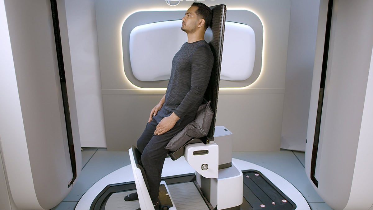
Since the beginning of radiation therapy, almost all treatments have been delivered with the patient lying on a table while the beam rotates around them. But a resurgence in upright patient positioning is changing that paradigm. Novel radiation accelerators such as proton therapy, VHEE, and FLASH therapy are often too large to rotate around the patient, making access limited. By instead rotating the patient, these previously hard-to-access beams could now become mainstream in the future.
Join leading clinicians and experts as they discuss how this shift in patient positioning is enabling exploration of new treatment geometries and supporting the development of advanced future cancer therapies.
L-R Serdar Charyyev, Eric Deutsch, Bill Loo, Rock Mackie
Novel beams covered and their representative speaker
Serdar Charyyev – Proton Therapy – Clinical Assistant Professor at Stanford University School of Medicine Eric Deutsch – VHEE FLASH – Head of Radiotherapy at Gustave Roussy Bill Loo – FLASH Photons – Professor of Radiation Oncology at Stanford Medicine Rock Mackie – Emeritus Professor at University of Wisconsin and Co-Founder and Chairman of Leo Cancer Care
This year saw Physics World report on a raft of innovative and exciting developments in the worlds of medical physics and biotech. These included novel cancer therapies using low-temperature plasma or laser ablation, intriguing new devices such as biodegradable bone screws and a pacemaker smaller than a grain of rice, and neural engineering breakthroughs including an ultrathin bioelectric implant that improves movement in rats with spinal cord injuries and a tiny brain sensor that enables thought control of external devices. Here are a few more research highlights that caught my eye.
Vision transformed
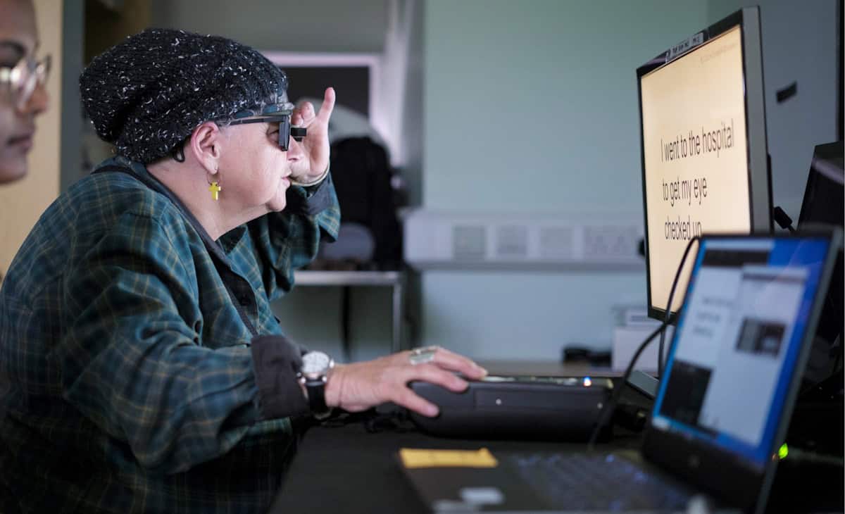
One remarkable device introduced in 2025 was an eye implant that restored vision to patients with incurable sight loss. In a clinical study headed up at the University of Bonn, participants with sight loss due to age-related macular degeneration had a tiny wireless implant inserted under their retina. Used in combination with specialized glasses, the system restored the ability to read in 27 of 32 participants followed up a year later.
Learning to read again Study participant Sheila Irvine, a patient at Moorfields Eye Hospital, training with the PRIMA device. (Courtesy: Moorfields Eye Hospital)
We also described a contact lens that enables wearers to see near-infrared light without night vision goggles, reported on an fascinating retinal stimulation technique that enabled volunteers to see colours never before seen by the human eye, and chatted with researchers in Hungary about how a tiny dissolvable eye insert they are developing could help astronauts suffering from eye conditions.
Radiation therapy advances
2025 saw several firsts in the field of radiation therapy. Researchers in Germany performed the first cancer treatment using a radioactive carbon ion beam, on a mouse with a bone tumour close to the spine. And a team at the Trento Proton Therapy Centre in Italy delivered the first clinical treatments using proton arc therapy – a development that made it onto our top 10 Breakthroughs of the Year.
Meanwhile, the ASTRO meeting saw Leo Cancer Care introduce its first upright photon therapy system, called Grace, which will deliver X-ray radiation to patients in an upright position. This new take on radiation delivery is also under investigation by a team at RaySearch Laboratories, who showed that combining static arcs and shoot-through beams could increase plan quality and reduce delivery times in upright proton therapy.
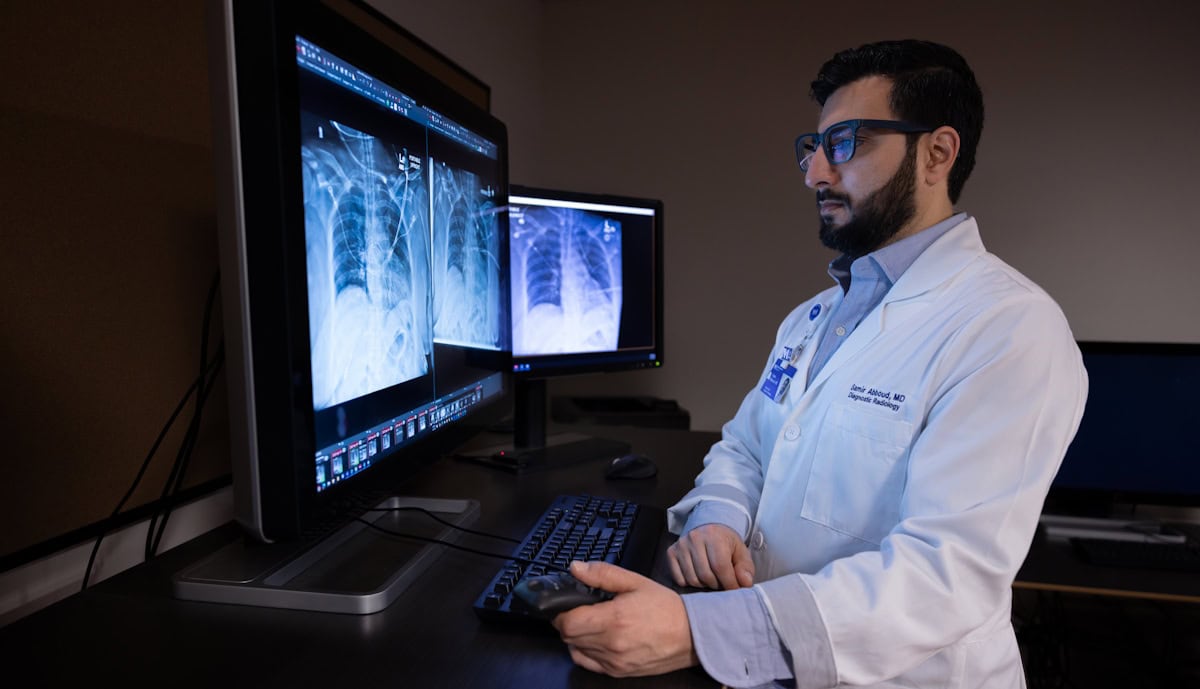
It’s particularly interesting to examine how the rapid evolution of artificial intelligence (AI) is impacting healthcare, especially considering its potential for use in data-intensive tasks. Earlier this year, a team at Northwestern Medicine integrated a generative AI tool into a live clinical workflow for the first time, using it to draft radiology reports on X-ray images. In routine use, the AI model increased documentation efficiency by an average of 15.5%, while maintaining diagnostic accuracy.
Samir Abboud: “For me and my colleagues, it’s not an exaggeration to say that [the AI tool] doubled our efficiency.” (Courtesy: José M Osorio/Northwestern Medicine)
When introducing AI into the clinic, however, it’s essential that any AI-driven software is accurate, safe and trustworthy. To help assess these factors, a multinational research team identified potential pitfalls in the evaluation of algorithmic bias in AI radiology models, suggesting best practices to mitigate such bias.
A quantum focus
Finally, with 2025 being the International Year of Quantum Science and Technology, Physics World examined how quantum physics looks set to play a key role in medicine and healthcare. Many quantum-based companies and institutions are already working in the healthcare sector, with quantum sensors, in particular, close to being commercialized. As detailed in this feature on quantum sensing, such technologies are being applied for applications ranging from lab and point-of-care diagnostics to consumer wearables for medical monitoring, body scanning and microscopy.
Alongside, scientists at Jagiellonian University are applying quantum entanglement to cancer diagnostics and developing the world’s first whole-body quantum PET scanner, while researchers at the University of Warwick have created an ultrasensitive magnetometer based on nitrogen-vacancy centres in diamond that could detect small cancer metastases via keyhole surgery. There’s even a team designing a protein qubit that can be produced directly inside living cells and used as a magnetic field sensor (which also featured in this year’s top 10 breakthroughs).
And in September, we ran a Physics World Live event examining how quantum optics, quantum sensors and quantum entanglement can enable advanced disease diagnostics and transform medical imaging. The recording is available to watch here.
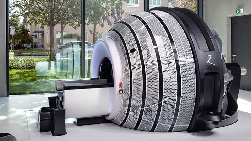
ZAP-X is a next-generation, cobalt-free, vault-free stereotactic radiosurgery system purpose-built for the brain. Delivering highly precise, non-invasive treatments with exceptionally low whole-brain and whole-body dose, ZAP-X’s gyroscopic beam delivery, refined beam geometry and fully integrated workflow enable state-of-the-art SRS without the burdens of radioactive sources or traditional radiation bunkers.
Theresa Hofman
Theresa Hofman is deputy head of medical physics at the European Radiosurgery Center Munich (ERCM), specializing in stereotactic radiosurgery with the CyberKnife and ZAP‑X systems. She has been part of the ERCM team since 2018 and has extensive clinical experience with ZAP‑X, one of the first centres worldwide to implement the technology in 2021. Since then, the team has treated more than 900 patients with ZAP‑X, and she is deeply involved in both clinical use and evaluation of its planning software.
She holds a master’s degree in physics from Ludwig Maximilian University of Munich, where she authored two first‑author publications on range verification in carbon‑ion therapy. At ERCM, she has published additional first‑author studies on CyberKnife kidney‑treatment accuracy and on comparative planning between ZAP‑X and CyberKnife. She is currently conducting further research on the latest ZAP‑X planning software. Her work is driven by the goal of advancing high‑quality radiosurgery and ensuring the best possible treatment for every patient.
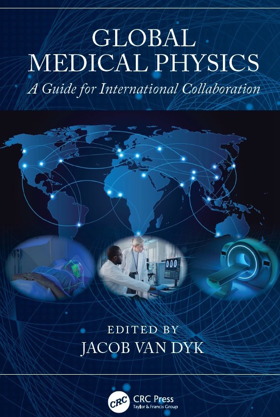
Educational aid Global Medical Physics: A Guide for International Collaboration explores the increasing role of medical physicists in international collaborations. The book comes in paperback, hardback and ebook format. An open-access ebook will be available in the near future. (Courtesy: CRC Press/Taylor & Francis)
As the world population ages and the incidence of cancer and cardiac disease grows alongside, there’s an ever-increasing need for reliable and effective diagnostics and treatments. Medical physics plays a central role in both of these areas – from the development of a suite of advanced diagnostic imaging modalities to the ongoing evolution of high-precision radiotherapy techniques.
But access to medical physics resources – whether equipment and infrastructure, education and training programmes, or the medical physicists themselves – is massively imbalanced around the world. In low- and middle-income countries (LMICs), fewer than 50% of patients have access to radiotherapy, with similar shortfalls in the availability of medical imaging equipment. Lower-income countries also have the least number of medical physicists per capita.
This disparity has led to an increasing interest in global health initiatives, with professional organizations looking to provide support to medical physicists in lower income regions. Alongside, medical physicists and other healthcare professionals seek to collaborate internationally in clinical, educational and research settings.
Successful multicultural collaborations, however, can be hindered by cultural, language and ethical barriers, as well as issues such as poor access to the internet and the latest technology advances. And medical physicists trained in high-income contexts may not always understand the circumstances and limitations of those working within lower income environments.
Aiming to overcome these obstacles, a new book entitled Global Medical Physics: A Guide for International Collaboration provides essential guidance for those looking to participate in such initiatives. The text addresses the various complexities of partnering with colleagues in different countries and working within diverse healthcare environments, encompassing clinical and educational medical physics circles, as well as research and academic environments.
“I have been involved in providing support to medical physicists in lower income contexts for a number of years, especially through the International Atomic Energy Agency (IAEA), but also through professional organizations like the American Association of Physicists in Medicine (AAPM),” explains the book’s editor Jacob Van Dyk, emeritus professor at Western University in Canada. “It is out of these experiences that I felt it might be appropriate and helpful to provide some educational materials that address these issues. The outcome was this book, with input from those with these collaborative experiences.”
Shared experience
The book brings together contributions from 34 authors across 21 countries, including both high- and low-resource settings. The authors – selected for their expertise and experience in global health and medical physics activities – provide guidelines for success, as well as noting potential barriers and concerns, on a wide range of themes targeted at multiple levels of expertise.
This guidance includes, for example: advice on how medical physicists can contribute to educational, clinical and research-based global collaborations and the associated challenges; recommendations on building global inter-institutional collaborations, covering administrative, clinical and technical challenges and ethical issues; and a case study on the Radiation Planning Assistant project, which aims to use automated contouring and treatment planning to assist radiation oncologists in LMICs.
In another chapter, the author describes the various career paths available to medical physicists, highlighting how they can help address the disparity in healthcare resources through their careers. There’s also a chapter focusing on CERN as an example of a successful collaboration engaging a worldwide community, including a discussion of CERN’s involvement in collaborative medical physics projects.
With the rapid emergence of artificial intelligence (AI) in healthcare, the book takes a look at the role of information and communication technologies and AI within global collaborations. Elsewhere, authors highlight the need for data sharing in medical physics, describing example data sharing applications and technologies.
Other chapters consider the benefits of cross-sector collaborations with industry, sustainability within global collaborations, the development of effective mentoring programmes – including a look at challenges faced by LMICs in providing effective medical physics education and training – and equity, diversity and inclusion and ethical considerations in the context of global medical physics.
The book rounds off by summarizing the key topics discussed in the earlier chapters. This information is divided into six categories: personal factors, collaboration details, project preparation, planning and execution, and post-project considerations.
“Hopefully, the book will provide an awareness of factors to consider when involved in global international collaborations, not only from a high-income perspective but also from a resource-constrained perspective,” says Van Dyk. “It was for this reason that when I invited authors to develop chapters on specific topics, they were encouraged to invite a co-author from another part of the world, so that it would broaden the depth of experience.”
 Since the beginning of radiation therapy, almost all treatments have been delivered with the patient lying on a table while the beam rotates around them. But a resurgence in upright patient positioning is changing that paradigm. Novel radiation accelerators such as proton therapy, VHEE, and FLASH therapy are often too large to rotate around the patient, making access limited. By instead rotating the patient, these previously hard-to-access beams could now become mainstream in the future.
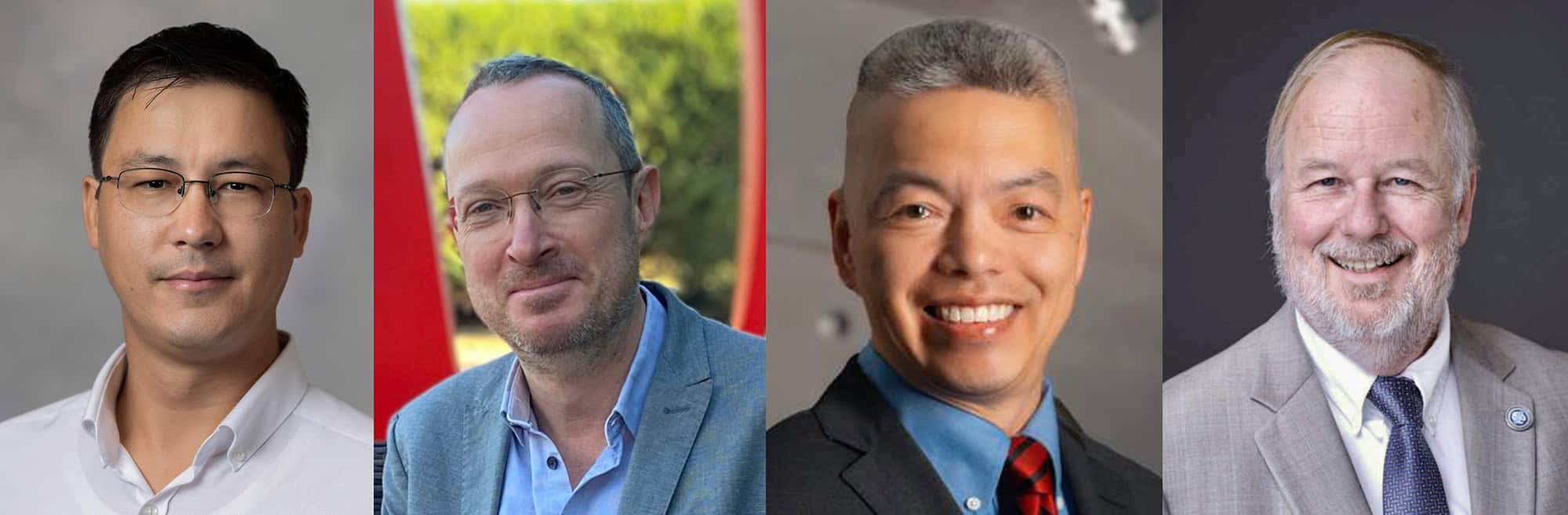
 ZAP-X is a next-generation, cobalt-free, vault-free stereotactic radiosurgery system purpose-built for the brain. Delivering highly precise, non-invasive treatments with exceptionally low whole-brain and whole-body dose, ZAP-X’s gyroscopic beam delivery, refined beam geometry and fully integrated workflow enable state-of-the-art SRS without the burdens of radioactive sources or traditional radiation bunkers.