↩ Accueil

Vue lecture

Il y a de nouveaux articles disponibles, cliquez pour rafraîchir la page.

Novel MRI technique can quantify lung function

Par : Tami Freeman

Assessing lung function is crucial for diagnosing and monitoring respiratory diseases. The most common way to do this is using spirometry, which measures the amount and speed of air that a person can inhale and exhale. Spirometry, however, is insensitive to early disease and cannot detect heterogeneity in lung function. Techniques such as chest radiography or CT provide more detailed spatial information, but are not ideal for long-term monitoring as they expose patients to ionizing radiation.

Now, a team headed up at Newcastle University in the UK has demonstrated a new lung MR imaging technique that provides quantitative and spatially localized assessment of pulmonary ventilation. The researchers also show that the MRI scans – recorded after the patient inhales a safe gas mixture – can track improvements in lung function following medication.

Although conventional MRI of the lungs is challenging, lung function can be assessed by imaging the distribution of an inhaled gas, most commonly hyperpolarized 3He or 129Xe. These gases can be expensive, however, and the magnetic preparation step requires extra equipment and manpower. Instead, project leader Pete Thelwall and colleagues are investigating 19F-MRI of inhaled perfluoropropane – an inert gas that does not require hyperpolarization to be visible in an MRI scan.

“Conventional MRI detects magnetic signals from hydrogen nuclei in water to generate images of water distribution,” Thelwall explains. “Perfluoropropane is interesting to us as we can also get an MRI signal from fluorine nuclei and visualize the distribution of inhaled perfluoropropane. We assess lung ventilation by seeing how well this MRI-visible gas moves into different parts of the lungs when it is inhaled.”

Testing the new technique

The researchers analysed 19F-MRI data from 38 healthy participants, 35 with asthma and 21 with chronic obstructive pulmonary disease (COPD), reporting their findings in Radiology. For the 19F-MRI scans, participants were asked to inhale a 79%/21% mixture of perfluoropropane and oxygen and then hold their breath. All subjects also underwent spirometry and an anatomical 1H-MRI scan, and those with respiratory disease withheld their regular bronchodilator medication before the MRI exams.

After co-registering each subject’s anatomical (1H) and ventilation (19F) images, the researchers used the perfluoropropane distribution in the images to differentiate ventilated and non-ventilated lung regions. They then calculated the ratio of non-ventilated lung to total lung volume, a measure of ventilation dysfunction known as the ventilation defect percentage (VDP).

Healthy subjects had a mean VDP of 1.8%, reflecting an even distribution of inhaled gas throughout their lungs and well-preserved lung function. In comparison, the patient groups showed elevated mean VDP values – 8.3% and 27.2% for those with asthma and COPD, respectively – reflecting substantial ventilation heterogeneity.

In participants with respiratory disease, the team also performed 19F-MRI after treatment with salbutamol, a common inhaler. They found that the MR images revealed changes in regional ventilation in response to this bronchodilator therapy.

Post-treatment images of patients with asthma showed an increase in lung regions containing perfluoropropane, reflecting the reversible nature of this disease. Participants with COPD generally showed less obvious changes following treatment, as expected for this less reversible disease. Bronchodilator therapy reduced the mean VDP by 33% in participants with asthma (from 8.3% to 5.6%) and by 14% in those with COPD (from 27.2% to 23.3%).

The calculated VDP values were negatively associated with standard spirometry metrics. However, the team note that some participants with asthma exhibited normal spirometry but an elevated mean VDP (6.7%) compared with healthy subjects. This finding suggests that the VDP acquired by 19F-MRI of inhaled perfluoropropane is more sensitive to subclinical disease than conventional spirometry.

Supporting lung transplants

In a separate study reported in JHLT Open, Thelwall and colleagues used dynamic 19F-MRI of inhaled perfluoropropane to visualize the function of transplanted lungs. Approximately half of lung transplant recipients experience organ rejection, known as chronic lung allograft dysfunction (CLAD), within five years of transplantation.
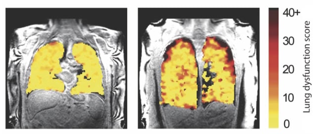
Lung function MRI
Early detection Lung function MRI showing areas of dysfunction in transplant recipients. (Courtesy: Newcastle University, UK)

Transplant recipients are monitored frequently using pulmonary function tests and chest X-rays. But by the time CLAD is diagnosed, irreversible lung damage may already have occurred. The team propose that 19F-MRI may find subtle early changes in lung function that could help detect rejection earlier.

The researchers studied 10 lung transplant recipients, six of whom were experiencing chronic rejection. They used a wash-in and washout technique, acquiring breath-hold 19F-MR images while the patient inhaled a perfluoropropane/oxygen mixture (wash-in acquisitions), followed by scans during breathing of room air (washout acquisitions).

The MR images revealed quantifiable differences in regional ventilation in participants with and without CLAD. In those with chronic rejection, scans showed poorer air movement to the edges of the lungs, likely due to damage to the small airways, a typical feature of CLAD. By detecting such changes in lung function, before signs of damage are seen in other tests, it’s possible that this imaging method might help inform patient treatment decisions to better protect the transplanted lungs from further damage.

The studies fall squarely within the field of clinical research, requiring non-standard MRI hardware to detect fluorine nuclei. But Thelwall sees a pathway towards introducing 19F-MRI in hospitals, noting that scanner manufacturers have brought systems to market that can detect nuclei other than 1H in routine diagnostic scans. Removing the requirement for hyperpolarization, combined with the lower relative cost of perfluoropropane inhalation (approximately £50 per study participant), could also help scale this method for use in the clinic.

The team is currently working on a study looking at how MRI assessment of lung function could help reduce the side effects associated with radiotherapy for lung cancer. The idea is to design a radiotherapy plan that minimizes dose to lung regions with good function, whilst maintaining effective cancer treatment.

“We are also looking at how better lung function measurements might help the development of new treatments for lung disease, by being able to see the effects of new treatments earlier and more accurately than current lung function measurements used in clinical trials,” Thelwall tells Physics World.

The post Novel MRI technique can quantify lung function appeared first on Physics World.
Magnetic particle imaging designed for the human brain

Par : Tami Freeman

Magnetic particle imaging (MPI) is an emerging medical imaging modality with the potential for high sensitivity and spatial resolution. Since its introduction back in 2005, researchers have built numerous preclinical MPI systems for small-animal studies. But human-scale MPI remains an unmet challenge. Now, a team headed up at the Athinoula A Martinos Center for Biomedical Imaging has built a proof-of-concept human brain-scale MPI system and demonstrated its potential for functional neuroimaging.

MPI works by visualizing injected superparamagnetic iron oxide nanoparticles (SPIONs). SPIONs exhibit a nonlinear response to an applied magnetic field: at low fields they respond roughly linearly, but at larger field strengths, particle response saturates. MPI exploits this behaviour by creating a magnetic field gradient across the imaging space with a field-free line (FFL) in the centre. Signals are only generated by the unsaturated SPIONs inside the FFL, which can be scanned through the imaging space to map SPION distribution.

First author Eli Mattingly and colleagues propose that MPI could be of particular interest for imaging the dynamics of blood volume in the brain, as it can measure the local distribution of nanoparticles in blood without an interfering background signal.

“In the brain, the tracer stays in the blood so we get an image of blood volume distribution,” Mattingly explains. “This is an important physiological parameter to map since blood is so vital for supporting metabolism. In fact, when a brain area is used by a mental task, the local blood volume swells about 20% in response, allowing us to map functional brain activity by dynamically imaging cerebral blood volume.”

Rescaling the scanner

The researchers began by defining the parameters required to build a human brain-scale MPI system. Such a device should be able to image the head with 6 mm spatial resolution (as used in many MRI-based functional neuroimaging studies) and 5 s temporal resolution for at least 30 min. To achieve this, they rescaled their existing rodent-sized imager.

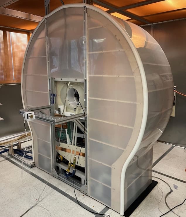
Human brain-scale MPI scanner
Proof-of-concept system The back of the MPI scanner showing the opening for the patient head. (Courtesy: Lawrence Wald)

The resulting scanner uses two opposed permanent magnets to generate the FFL and high-power electromagnet shift coils, comprising inner and outer coils on each side of the head, to sweep the FFL across the head. The magnets create a gradient of 1.13 T/m, sufficient to achieve 5–6 mm resolution with high-performance SPIONs. To create 2D images, a mechanical gantry rotates the magnets and shift coils at 6 RPM, enabling imaging every 5 s.

The MPI system also incorporates a water-cooled 26.3 kHz drive coil, which produces the oscillating magnetic field (of up to 7 mTpeak) needed to drive the SPIONs in and out of saturation. A gradiometer-based receive coil fits over the head to record the SPION response.

Mattingly notes that this rescaling was far from straightforward as many parameters scale with the volume of the imaging bore. “With a bore about five times larger, the volume is about 125 times larger,” he says. “This means the power electronics require one to two orders of magnitude more power than rat-sized MPI systems, and the receive coils are simultaneously less sensitive as they become larger.”

Performance assessment

The researchers tested the scanner performance using a series of phantoms. They first evaluated spatial resolution by imaging 2.5 mm-diameter capillary tubes filled with Synomag SPIONs and spaced by between 5 and 9 mm. They reconstructed images using an inverse Radon reconstruction algorithm and a forward-model iterative reconstruction.

The system demonstrated a spatial resolution of about 7 mm with inverse Radon reconstruction, increasing to 5 mm with iterative reconstruction. The team notes that this resolution should be sufficient to observe changes in cerebral blood volume associated with brain function and following brain injuries.

To determine the practical detection limit, the researchers imaged Synomag samples with concentrations from 6 mgFe/ml to 15.6 µgFe/ml, observing a limit of about 1 µgFe. Based on this result, they predict that MPI should show grey matter with a signal-to-noise ratio (SNR) of roughly five and large blood vessels with an SNR of about 100 in a 5 s image. They also expect to detect changes during brain activation with a contrast-to-noise ratio of above one.

Next, they quantified the scanner’s imaging field-of-view using a G-shaped phantom filled with Synomag at roughly the concentration of blood. The field-of-view was 181 mm in diameter – sufficient to encompass most human brains. Finally, the team monitored the drive current stability over 35 min of continuous imaging. At a drive field of 4.6 mTpeak, the current deviated less than 2%. As this drift was smooth and slow, it should be straightforward to separate it from the larger signal changes expected from brain activation.

The researchers conclude that their scanner – the first human head-sized, mechanically rotating, FFL-based MPI – delivers a suitable spatial resolution, temporal resolution and sensitivity for functional human neuroimaging. And they continue to improve the device. “Currently, the group is developing hardware to enable studies such as application-specific receive coils to prepare for in vivo experiments,” says Mattingly.

At present, the scanner’s sensitivity is limited by background noise from the amplifiers. Mitigating such noise could increase sensitivity 20-fold, the team predicts, potentially providing an order of magnitude improvement over other human neuroimaging methods and enabling visualization of haemodynamic changes following brain activity.

The MPI system is described in Physics in Medicine & Biology.

The post Magnetic particle imaging designed for the human brain appeared first on Physics World.

Imaging and medical-physics firms bag Institute of Physics business awards 2024

In my previous article, I highlighted some of the quantum and green-energy companies that won Business Innovation Awards from the Institute of Physics in 2024. But imaging and medical-physics firms did well too. Having sat on the judging panel for the awards, I saw some fantastic entries – and picking winners wasn’t easy. Let me start, though, with Geoptic, which is one of an elite group of firms to win a second IOP business award, adding a Business Innovation Award to its start-up prize in 2020.

Geoptic is a spin-out from three collaborating groups of physicists at the universities of Durham, Sheffield and St Mary’s Twickenham. The company uses cosmic-ray muon radiography and tomography to study large engineering structures. In particular, it was honoured by the IOP for using the technique to ensure the safety of tunnels on the UK’s railway network.

Many of the railway tunnels in the UK date back to the mid-19th century. To speed up construction, temporary shafts were bored vertically down below the ground, allowing workers to dig at multiple points along the route of the tunnel. When the tunnel was complete, the shafts would be sealed, but their precise number and location is often unclear.

The shafts are a major hazard to the tunnel’s integrity, which is not great for Network Rail – the state-owned body that’s responsible for the UK’s rail infrastructure. Geoptic has, however, been working with Network Rail to provide its engineers with a clear structural view of the dangers that lurk along its route. In my view, it’s a really innovating imaging company, solving challenging real-world problems.

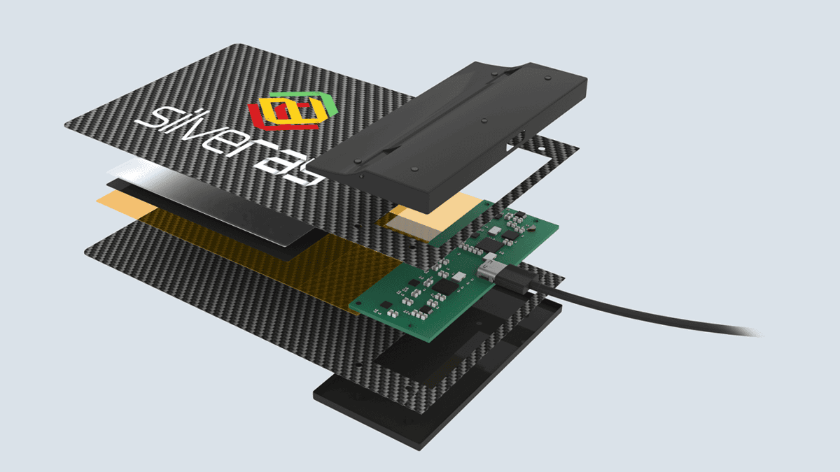
Another winner is Silveray, which was spun off from the University of Surrey. It’s picked up an IOP Business Start-up Award for creating flexible, “colour” X-ray detectors based on proprietary semiconductor materials. Traditional X-ray images are black and white, but what Silveray has done is to develop a nano-particle semiconductor ink that can be coated on to any surface and work at multiple wavelengths.

Silveray's digital X-ray film
Visionary idea Silveray won an IOP Business Start-up Award for creating flexible, “colour” X-ray detectors based on proprietary semiconductor materials. (Courtesy: Silveray)

The X-ray detectors, which are flexible, can simply be wrapped around pipes and other structures that need to be imaged. Traditionally, this has been done using analogue X-ray film that has to be developed in an off-site dark room. That’s costly and time-consuming – especially if images failed to be recorded. Silveray’s detectors instead provide digital X-ray images in real time, making it an exciting and innovative technology that could transform the $5bn X-ray detector market.

Phlux Technology, meanwhile, has won an IOP Business Start-up Award for developing patented semiconductor technology for infrared light sensors that are 12 times more sensitive than the best existing devices, making them ideal for fast, accurate 3D imaging. Set up by researchers at the University of Sheffield, Phlux’s devices have many potential applications especially in light detection and ranging (LIDAR), laser range finders, optical-fibre test instruments and optical and quantum communications networks.

In LIDAR, Phlux’s can have 12 times greater image resolution for a given transmitter power. Its sensors could also make vehicles much safer by enabling higher-resolution images to be created over longer distances, making safety systems more effective. The first volume market for the company is likely to be in communications and where a >10 dB increase in detector sensitivity is going to be well received by the market.

Given the number of markets that will benefit from an “over an order of magnitude” improvement, Phlux is one to watch for a future Business Innovation Award too.

Medical marvel

Let me finish by mentioning Crainio, a medical technology spin-off company from City, University of London, which has won the 2024 Lee Lucas award. This award honours promising start-up firms in the medical and healthcare sector thanks to a generous donation by Mike and Ann Lee (née Lucas). These companies need all the support, time and money they can get given the many challenging regulatory requirements in the medical sector.

Crainio’s technology allows healthcare workers to measure intracranial pressure (ICP), a vital indicator of brain health after a head injury. Currently, the only way to measure ICP directly is for a neurosurgeon to drill a hole in a patient’s skull and place an expensive probe in the brain. It’s a highly invasive procedure that can’t easily be carried out in the “golden hours” immediately after an accident, requiring access to scarce and expensive neurosurgery resources. The procedure is also medically risky, leading to potential infection, bleeding and other complications.

Crainio’s technology eliminates these risks, enabling direct measurement of ICP through a simple non-invasive probe applied to the forehead. The technology – using infrared photoplethysmography (PPG) combined with machine learning – is based on years of research and development work conducted by Panicos Kyriacou and his team of biomedical engineers at City.

Good levels of accuracy have been demonstrated in clinical studies conducted at the Royal London Hospital. It certainly seems a much better plan than drilling a hole in your head as I am sure you can agree – making Crainio a worthy winner, with its non-invasive technology it should have a positive impact on patients globally. I hope the regulatory hurdles can be quickly cleared so the company can start helping patients as soon as possible.

As I have mentioned before, all physics-based firms require time and energy to develop products and become globally significant. There’s also the perennial difficulty of explaining a product idea, which is often quite specialized, to potential investors who have little or no science background. An IOP start-up award can therefore show that your technology has won approval from judges with solid physics and business experience.

I hope, therefore, that your company, if you have one, will be inspired to apply. Also remember that the IOP offers three other awards (Katharine Burr Blodgett, Denis Gabor and Clifford Paterson) for individuals or teams who have been involved in innovative physics with a commercial angle. Good luck – and remember, you have to be in it to win it. Award entries for 2025 will be open in February 2025.

The post Imaging and medical-physics firms bag Institute of Physics business awards 2024 appeared first on Physics World.

AI-powered tool detects residual tumour during brain surgery

Par : No Author
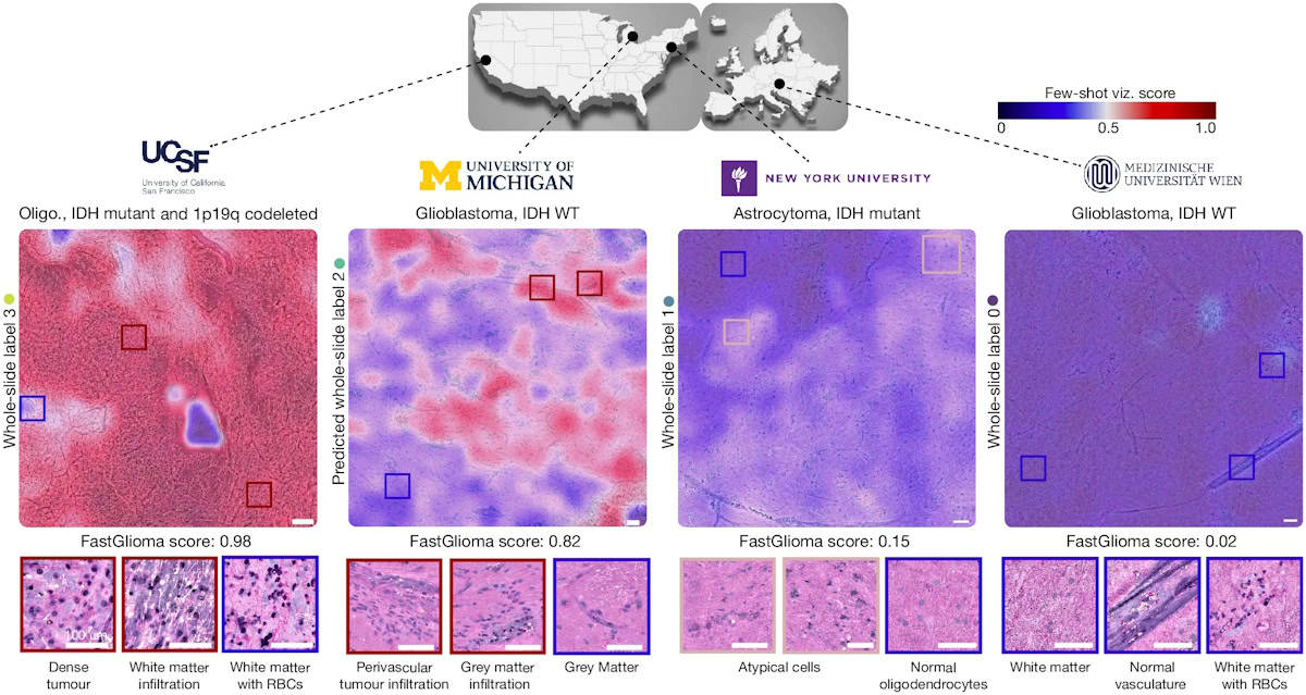
The FastGlioma diagnostic tool
Fast and accurate FastGlioma is an artificial intelligence-based diagnostic system that can detect residual brain tumour that’s often missed during surgery. The image shows various diffuse glioma specimens from the four study centres. Scale bars, 100 μm. (Courtesy: Nature 10.1038/s41586-024-08169-3)

When surgery is performed to remove cancerous tissue, one question always lingers: “did the surgeon get everything?”.

In the case of brain tumour resection, the answer is often “no”. Residual cancerous tissue at the edges of a cavity where a malignant mass has been removed can visually resemble healthy tissue and be overlooked, or may be microscopic in size.

A new tool for neurosurgeons, designed for fast and accurate detection of microscopic brain tumour infiltration in unprocessed tissue samples from surgical margins, may lead to a new era of success for brain cancer surgery.

The developers of the new FastGlioma tool, at the University of Michigan and the University of California, San Francisco (UCSF), explain that it can predict if and the extent to which glioma remains in the brain while the surgical procedure is underway. FastGlioma also provides visual heat-map guidance of the location(s) requiring additional reaction for safe maximal tumour removal.

FastGlioma combines rapid, easy-to-use stimulated Raman histology (SRH) optical imaging with open-source visual foundation models (artificial intelligence models trained on massive, diverse datasets that can be adapted for a wide range of tasks) to perform a 10 s analysis of fresh tissue specimens in operating room suites. FastGlioma proved not only significantly faster and cheaper than conventional standard-of-care MRI- and fluorescence-based surgical guidance, but in head-to-head comparisons, it significantly outperformed them in detection of two types of glioma (IDH wild-type and IDH-mutant diffuse gliomas).

Training and validation

In a prospective multicentre clinical study, principal investigators Todd Hollon of the University of Michigan and Shawn Hervey-Jumper from UCSF and co-researchers trained and validated FastGlioma to detect microscopic tumour infiltration in an international cohort of patients. They explain that “foundation modelling had not been previously investigated in studies on the clinical applications of SRH”, adding that they focused on tumour infiltration “as the most clinically important and ubiquitous problem in cancer surgery”.

The researchers trained FastGlioma using 11,462 whole-slide SRH images, divided into around four million unique 300×300 pixel SRH patches, acquired from 2799 patients undergoing surgery for suspected central nervous system tumours and/or epilepsy. They validated the model using a dataset of 3560 whole-slide images (852,000 patches) from 896 patients. Diagnostic classes of the dataset included normal brain, high-grade glioma, low-grade glioma, meningioma, pituitary adenoma, schwannoma and metastatic tumour. A subset of these had tumour infiltration categorized, ranging from normal brain to dense infiltration.

The researchers also developed a rapid visualization strategy, called few-shot visualizations. Based on FastGlioma’s self-supervised training, few-shot visualizations use a small support set of physician-selected SRH patch examples, representing a diverse selection of diffuse gliomas and normal brain parenchyma. By comparing feature similarity between the support set and the tissue sample being analysed, FastGlioma creates both a tumour-infiltration score and infiltration heat maps.

Prospective clinical testing

To test the fine-tuned FastGlioma model, three medical centres – UCSF, NYU Langone in New York City and the Medical University of Vienna – enrolled 220 patients with suspected diffuse gliomas who underwent tumour resection.

FastGlioma could detect and quantify the degree of tumour infiltration with an average accuracy of 92.1%. The tool maintained accurate tumour-infiltration scores despite significant cytological and histoarchitectural differences related to tumour grade, molecular genetics, treatment effect or WHO subtypes.

The primary end point for the study, reported in Nature, was to validate the accuracy and reproducibility of FastGlioma across various patient populations, demographics, medical centres and World Health Organization (WHO) diffuse glioma molecular subgroups. Additionally, the team aimed to compare the performance of FastGlioma with standard-of-care methods for intraoperative tumour-infiltration detection during brain tumour surgery.

To achieve this, the researchers evaluated FastGlioma as a surgical adjunct in a subset of 129 patients. Neurosurgeons sampled surgical margins at their discretion. Following SHR-imaging during the surgical procedure, the resected specimens were preserved for subsequent microscopic analysis. Expert neuropathologists scored each SRH image postoperatively to provide ground truth tumour-infiltration scores. FastGlioma significantly outperformed conventional methods, with only a 3.8% tumour miss rate, compared with a 24% miss rate for current standard-of-care surgical guidance methods.

Another benefit is that the analytic speed of FastGlioma provides a rapid and scalable alternative to conventional intraoperative pathology methods. The researchers point out that visual foundation models like FastGlioma also minimize reliance on radiographic features, contrast enhancement or extrinsic fluorescent labels to optimize the extent of resection.

The researchers also note that FastGlioma can accurately detect residual tumour for several non-glioma brain tumours, including paediatric brain tumours. “FastGlioma represents the transformative potential of medical foundation models to unlock the role of artificial intelligence in care of patients with cancer,” they write.

Future research will focus on applying a similar workflow to other human cancers, including lung, prostate, head-and-neck and breast cancer.

The post AI-powered tool detects residual tumour during brain surgery appeared first on Physics World.

New imaging technique could change how we look at certain objects in space

A new imaging technique that takes standard two-dimensional (2D) radio images and reconstructs them as three-dimensional (3D) ones could tell us more about structures such as the jet-like features streaming out of galactic black holes. According to the technique’s developers, it could even call into question physical models of how radio galaxies formed in the first place.

“We will now be able to obtain information about the 3D structures in polarized radio sources whereas currently we only see their 2D structures as they appear in the plane of the sky,” explains Lawrence Rudnick, an observational astrophysicist at the University of Minnesota, US, who led the study. “The analysis technique we have developed can be performed not only on the many new maps to be made with powerful telescopes such as the Square Kilometre Array and its precursors, but also from decades of polarized maps in the literature.”

Analysis of data from the MeerKAT radio telescope array

In their new work, Rudnick and colleagues in Australia, Mexico, the UK and the US studied polarized light data from the MeerKAT radio telescope array at the South African Radio Astronomy Observatory. They exploited an effect called Faraday rotation, which rotates the angle of polarized radiation as it travels through a magnetized ionized region. By measuring the amount of rotation for each pixel in an image, they can determine how much material that radiation passed through.

In the simplest case of a uniform medium, says Rudnick, this information tells us the relative distance between us and the emitting region for that pixel. “This allows us to reconstruct the 3D structure of the radiating plasma,” he explains.

An indication of the position of the emitting region

The new study builds on a previous effort that focused on a specific cluster of galaxies for which the researchers already had cubes of data representing its 2D appearance in the sky, plus a third axis given by the amount of Faraday rotation. In the latest work, they decided to look at this data in a new way, viewing the cubes from different angles.

“We realized that the third axis was actually giving us an indication of the position of the emitting region,” Rudnick says. “We therefore extended the technique to situations where we didn’t have cubes to start with, but could re-create them from a pair of 2D images.”

There is a problem, however, in that polarization angle can also rotate as the radiation travels through regions of space that are anything but uniform, including our own Milky Way galaxy and other intervening media. “In that case, the amount of radiation doesn’t tell us anything about the actual 3D structure of the emitting source,” Rudnick adds. “Separating out this information from the rest of the data is perhaps the most difficult aspect of our work.”

Shapes of structures are very different in 3D

Using this technique, Rudnick and colleagues were able determine the line-of-sight orientation of active galactic nuclei (AGN) jets as they are expelled from a massive black hole at the centre of the Fornax A galaxy. They were also able to observe how the materials in these jets interact with “cosmic winds” (essentially larger-scale versions of the magnetic solar wind streaming from our own Sun) and other space weather, and to analyse the structures of magnetic fields inside the jets from the M87 galaxy’s black hole.

The team found that the shapes of structures as inferred from 2D radio images were sometimes very different from those that appear in the 3D reconstructions. Rudnick notes that some of the mental “pictures” we have in our heads of the 3D structure of radio sources will likely turn out to be wrong after they are re-analysed using the new method. One good example in this study was a radio source that, in 2D, looks like a tangled string of filaments filling a large volume. When viewed in 3D, it turns out that these filamentary structures are in fact confined to a band on the surface of the source. “This could change the physical models of how radio galaxies are formed, basically how the jets from the black holes in their centres interact with the surrounding medium,” Rudnick tells Physics World.

The work is detailed in the Monthly Notices of the Royal Astronomical Society

The post New imaging technique could change how we look at certain objects in space appeared first on Physics World.

Cascaded crystals move towards ultralow-dose X-ray imaging

Par : Tami Freeman
Single-crystal and cascade-connected devices under X-ray irradiation
Working principle Illustration of the single-crystal (a) and cascade-connected two-crystal (b) devices under X-ray irradiation. (c) Time-resolved photocurrent responses of the two devices. (Courtesy: CC BY 4.0/ACS Cent. Sci. 10.1021/acscentsci.4c01296)

X-ray imaging plays an indispensable role in diagnosing and staging disease. Nevertheless, exposure to high doses of X-rays has potential for harm, and much effort is focused towards reducing radiation exposure while maintaining diagnostic function. With this aim, researchers at the King Abdullah University of Science and Technology (KAUST) have shown how interconnecting single-crystal devices can create an X-ray detector with an ultralow detection threshold.

The team created devices using lab-grown single crystals of methylammonium lead bromide (MAPbBr3), a perovskite material that exhibits considerable stability, minimal ion migration and a high X-ray absorption cross-section – making it ideal for X-ray detection. To improve performance further, they used cascade engineering to connect two or more crystals together in series, reporting their findings in ACS Central Science.

X-rays incident upon a semiconductor crystal detector generate a photocurrent via the creation of electron–hole pairs. When exposed to the same X-ray dose, cascade-connected crystals should exhibit the same photocurrent as a single-crystal device (as they generate equal net concentrations of electron–hole pairs). The cascade configuration, however, has a higher resistivity and should thus have a much lower dark current, improving the signal-to-noise ratio and enhancing the detection performance of the cascade device.

To test this premise, senior author Omar Mohammed and colleagues grew single crystals of MAPbBr3. They first selected four identical crystals to evaluate (SC1, SC2, SC3 and SC4), each 3 x 3 mm in area and approximately 2 mm thick. Measuring various optical and electrical properties revealed high consistency across the four samples.

“The synthesis process allows for reproducible production of MAPbBr3 single crystals, underscoring their strong potential for commercial applications,” says Mohammed.

Optimizing detector performance

Mohammed and colleagues fabricated X-ray detectors containing a single MAPbBr3 perovskite crystal (SC1) and detectors with two, three and four crystals connected in series (SC1−2, SC1−3 and SC1−4). To compare the dark currents of the devices they irradiated each one with X-rays under a constant 2 V bias voltage. The cascade-connected SC1–2 exhibited a dark current of 7.04 nA, roughly half that generated by SC1 (13.4 nA). SC1–3 and SC1–4 reduced the dark current further, to 4 and 3 nA, respectively.

The researchers also measured the dark current for the four devices as the bias voltage changed from 0 to -10 V. They found that SC1 reached the highest dark current of 547 nA, while SC1–2, SC1–3 and SC1–4 showed progressively decreasing dark currents of 134, 90 and 50 nA, respectively. “These findings highlight the effectiveness of cascade engineering in reducing dark current levels,” Mohammed notes.

Next, the team assessed the current stability of the devices under continuous X-ray irradiation for 450 s. SC1–2 exhibited a stable current response, with a skewness value of just 0.09, while SC1, SC1–3 and SC1–4 had larger skewness values of 0.75, 0.45 and 0.76, respectively.

The researchers point out that while connecting more single crystals in series reduced the dark current, increasing the number of connections also lowered the stability of the device. The two-crystal SC1–2 represents the optimal balance.

Low-dose imaging

One key component required for low-dose X-ray imaging is a low detection threshold. The conventional single-crystal SC1 showed a detection limit of 590 nGy/s under a 2 V bias. SC1–2 decreased this limit to 100 nGy/s – the lowest of all four devices and surpassing the existing record achieved by MAPbBr3 perovskite devices under near-identical conditions.

Spatial resolution is another important consideration. To assess this, the researchers estimated the modulation transfer function (the level of original contrast maintained by the detector) for each of the four devices. They found that SC1–2 exhibited the best spatial resolution of 8.5 line pairs/mm, compared with 5.6, 5.4 and 4 line pairs/mm for SC1, SC1–3 and SC1–4, respectively.

X-ray images of a key and a raspberry with a needle
Optimal imaging Actual and X-ray images of a key and a raspberry with a needle obtained by the SC1 to SC1–4 devices. (Courtesy: CC BY 4.0/ACS Cent. Sci. 10.1021/acscentsci.4c01296)

Finally, the researchers performed low-dose X-ray imaging experiments using the four devices, first imaging a key at a dose rate of 3.1 μGy/s. SC1 exhibited an unclear image due to the unstable current affecting its resolution. Devices SC1–2 to SC1–4 produced clearer images of the key, with SC1–2 showing the best image contrast.

They also imaged a USB port at a dose rate of 2.3 μGy/s, a metal needle piercing a raspberry at 1.9 μGy/s and an earring at 750 nGy/s. In all cases, SC1–2 exhibited the highest quality image.

The researchers conclude that the cascade-engineered configuration represents a significant shift in low-dose X-ray detection, with potential to advance applications that require minimal radiation exposure combined with excellent image quality. They also note that the approach works with different materials, demonstrating X-ray detection using cascaded cadmium telluride (CdTe) single crystals.

Mohammed says that the team is now investigating the application of the cascade structure in other perovskite single crystals, such as FAPbI3 and MAPbI3, with the goal of reducing their detection limits. “Moreover, efforts are underway to enhance the packaging of MAPbBr3 cascade single crystals to facilitate their use in dosimeter detection for real-world applications,” he tells Physics World.

The post Cascaded crystals move towards ultralow-dose X-ray imaging appeared first on Physics World.

From melanoma to malaria: photoacoustic device detects disease without taking a single drop of blood

Par : Tami Freeman

Malaria remains a serious health concern, with annual deaths increasing yearly since 2019 and almost half of the world’s population at risk of infection. Existing diagnostic tests are less than optimal and all rely on obtaining an invasive blood sample. Now, a research collaboration from USA and Cameroon has demonstrated a device that can non-invasively detect this potentially deadly infection without requiring a single drop of blood.

Currently, malaria is diagnosed using optical microscopy or antigen-based rapid diagnostic tests, but both methods have low sensitivity. Polymerase chain reaction (PCR) tests are more sensitive, but still require blood sampling. The new platform – Cytophone – uses photoacoustic flow cytometry (PAFC) to rapidly identify malaria-infected red blood cells via a small probe placed on the back of the hand.

PAFC works by delivering low-energy laser pulses through the skin into a blood vessel and recording the thermoacoustic signals generated by absorbers in circulating blood. Cytophone, invented by Vladimir Zharov from the University of Arkansas for Medical Science, was originally developed as a universal diagnostic platform and first tested clinically for detection of cancerous melanoma cells.

“We selected melanoma because of the possibility of performing label-free detection of circulating cells using melanin as an endogenous biomarker,” explains Zharov. “This avoids the need for in vivo labelling by injecting contrast agents into blood.” For malaria diagnosis, Cytophone detects haemozoin, an iron crystal that accumulates in red blood cells infected with malaria parasites. These haemozoin biocrystals have unique magnetic and optical properties, making them a potential diagnostic target.

Photoacoustic detection
Photoacoustic detection Schematic of the focused ultrasound transducer array assessing a blood network. (Courtesy: Nat. Commun. 10.1038/s41467-024-53243-z)

“The similarity between melanin and haemozoin biomarkers, especially the high photoacoustic contrast above the blood background, motivated us to bring a label-free malaria test with no blood drawing to malaria-endemic areas,” Zharov tells Physics World. “To build a clinical prototype for the Cameroon study we used a similar platform and just selected a smaller laser to make the device more portable.”

The Cytophone prototype uses a 1064 nm laser with a linear beam shape and a high pulse rate to interrogate fast moving blood cells within blood vessels. Haemozoin nanocrystals in infected red blood cells absorb this light (more strongly than haemoglobin in normal red blood cells), heat up and expand, generating acoustic waves. These signals are detected by an array of 16 tiny ultrasound transducers in acoustic contact with the skin. The transducers have focal volumes oriented in a line across the vessel, which increases sensitivity and resolution, and simplifies probe navigation.

In vivo testing

Zharov and collaborators – also from Yale School of Public Health and the University of Yaoundé I – tested the Cytophone in 30 Cameroonian adults diagnosed with uncomplicated malaria. They used data from 10 patients to optimize device performance and assess safety. They then performed a longitudinal study in the other 20 patients, who attended four or five times at up to 37 days following antimalarial therapy, contributing 94 visits in total.

Photoacoustic waveforms and traces from infected blood cells have a particular shape and duration, and a different time delay to that of background skin signals. The team used these features to optimize signal processing algorithms with appropriate averaging, filtration and gating to identify true signals arising from infected red blood cells. As the study subjects all had dark skin with high melanin content, this time-resolved detection also helped to avoid interference from skin melanin.

On visit 1 (the day of diagnosis), 19/20 patients had detectable photoacoustic signals. Following treatment, these signals consistently decreased with each visit. Cytophone-positive samples exhibited median photoacoustic peak rates of 1.73, 1.63, 1.18 and 0.74 peaks/min on visits 1–4, respectively. One participant had a positive signal on visit 5 (day 30). The results confirm that Cytophone is sensitive enough to detect low levels of parasites in infected blood.

The researchers note that Cytophone detected the most common and deadliest species of malaria parasite, as well as one infection by a less common species and two mixed infections. “That was a really exciting proof-of-concept with the first generation of this platform,” says co-lead author Sunil Parikh in a press statement. “I think one key part of the next phase is going to involve demonstrating whether or not the device can detect and distinguish between species.”

The research team
Team work The researchers from the USA and Cameroon are using photoacoustic flow cytometry to rapidly detect malaria infection. (Courtesy: Sunil Parikh)

Performance comparison

Compared with invasive microscopy-based detection, Cytophone demonstrated 95% sensitivity at the first visit and 90% sensitivity during the follow-up period, with 69% specificity and an area under the ROC curve of 0.84, suggesting excellent diagnostic performance. Cytophone also approached the diagnostic performance of standard PCR tests, with scope for further improvement.

Staff required just 4–6 h of training to operate Cytophone, plus a few days experience to achieve optimal probe placement. And with minimal consumables required and the increasing affordability of lasers, the researchers estimate that the cost per malaria diagnosis will be low. The study also confirmed that the safety of the Cytophone device. “Cytophone has the potential to be a breakthrough device allowing for non-invasive, rapid, label-free and safe in vivo diagnosis of malaria,” they conclude.

The researchers are now performing further malaria-related clinical studies focusing on asymptomatic individuals and children (for whom the needle-free aspect is particularly important). Simultaneously, they are continuing melanoma trials to detect early-stage disease and investigating the use of Cytophone to detect circulating blood clots in stroke patients.

“We are integrating multiple innovations to further enhance Cytophone’s sensitivity and specificity,” says Zharov. “We are also developing a cost-effective wearable Cytophone for continuous monitoring of disease progression and early warning of the risk of deadly disease.”

The study is described in Nature Communications.

The post From melanoma to malaria: photoacoustic device detects disease without taking a single drop of blood appeared first on Physics World.

❌