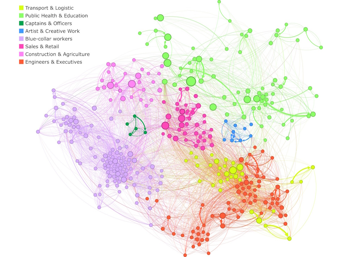
Occupational transition network Graphical visualization of the weighted and directed labour market network in France derived from the transition probability matrix, computed from data spanning 2012 to 2020. Each node symbolizes an occupation, with links illustrating transitions between them. Node sizes correspond to the occupation’s workforce size, line widths are proportional to the transition probability. (Courtesy: M Knicker, K Naumann-Woleske and M Benzaquen, École Polytechnique Paris)
A new statistical physics analysis of the French labour market has revealed that the vast majority of occupations act as so-called condenser occupations. These structural bottlenecks attract workers from many other types of jobs, but offer very limited options for further mobility. This finding could help explain why changing jobs in response to shocks like technological change or economic crises is often so slow, say scientists at the Ecole Polytechnique in Paris, who performed the study.
“By pinpointing where mobility gets ‘stuck’, we provide a new lens to understand – and potentially improve – the adaptability of labour markets,” explains Max Knicker of the EconophysiX lab, who led this new research effort.
Knicker and colleagues borrowed a concept from statistical physics known as the “fitness and complexity” framework, which is used to study the structure of economies and ecosystems. In their work, the researchers treated occupations as nodes in a network and analysed real transition data – that is, how workers actually moved between different jobs in France from 2012 to 2020. The data came from official sources and were provided by the National Institute of Statistics and Economic Studies through the Secure Data Access Center (CASD).
“In total, we had access to information on about 30 million workers and employers in France, whom we tracked over a 10-year period,” explains Knicker. “We also worked with high-resolution administrative data from INSEE (the French National Institute of Statistics), and specifically the BTS-Postes (Base Tous Salariés-Postes).”
Two key metrics
The researchers assigned a score for each occupation and developed two key metrics. These were: accessibility, which measures how many different jobs “feed into” a given occupation; and transferability, which measures how many different jobs someone can move to from that occupation.
By studying the network of job flows with these metrics, they observed hidden patterns and constraints in occupational mobility and identified four main clusters, or categories, of jobs. The first are defined as “diffuser” occupations and have high transferability but low accessibility. “These require specific training to enter, but that training allows for transitions to many other areas,” explains Knicker. “This means they are more difficult to get into, but offer a wide range of exit opportunities.”
The second group are called “channel” occupations. These are both hard to enter and offer few onward transitions, he says. “They often involve highly specialized skills, such as specific types of machine operation.”
The third class are “hubs” and are both widely accessible and highly transferable – so much so that they act as central nodes in the transition network. “This class includes jobs like retail sellers, which require a broad, yet not highly specialized skill set,” says Knicker.
The fourth and last category is the most common type and dubbed “condenser” occupations. “Workers from many different backgrounds can easily enter these, but they can’t easily get out afterwards,” explains Knicker. “Examples of such jobs include caregiving roles.”
A valuable tool for policymakers
The researchers explain that they undertook their study to answer a broader question: why do some economies adapt quickly to shocks while others struggle? “Despite increasing attention to issues like automation or the green transition, we still lacked tools to diagnose where worker mobility breaks down,” says Knicker. “A key challenge was dealing with the sheer complexity and size of the labour flow data – we analysed over 250 million person–year observations. Another was interpreting the results in a meaningful, policy-relevant way, since the transition network is shaped by many intertwined factors like skill compatibility, employer preferences and worker choices.”
The new framework could become a valuable tool for policymakers seeking to make labour markets more responsive, he tells Physics World. “For example, by identifying specific occupations that function as bottlenecks, we can better target reskilling efforts or job transition programmes. It also suggests that simply increasing training isn’t enough – what matters is where people are coming from and where they can go next.”
The researchers also showed that the structure of job transitions itself can limit mobility. Over time, this could inform the design of more strategic labour interventions, especially in the face of structural shocks like AI-driven job displacement, states Knicker.
Looking forward, the Ecole Polytechnique team plans to extend its approach by studying how the career paths of individual workers evolve over time. This, says Knicker, will be done using panel data, not just year-to-year snapshots as in the present analysis. He and his colleagues are also interested in linking their metrics to wage dynamics – for example, does low transferability make workers more vulnerable to exploitation or wage stagnation? “Finally, we hope to explore whether similar bottleneck structures exist in other countries, which could reveal whether these patterns are universal or country-specific.”
President Trump’s recent bombing of Iran’s nuclear sites marks a major turning point in the maintenance of global security. With war again erupting in the Middle East, the United States […]
Slam Corp, the shell company founded by former MLB player Alex Rodriguez, is suing Lynk Global to stop the direct-to-smartphone satellite operator from walking away from their long-delayed merger.
What does quantum physics have to do with vibrant oil paintings and the ghostly grin of a disappearing cat? Quite a lot, as it turns out. In this month’s Physics World Stories podcast, host Andrew Glester takes a colourful look at how we visualize – and try to make sense of – the curious world of quantum mechanics.
Felicity shares her journey from academia to art, and how her experience of number-colour synaesthesia – where numbers are associated with colours in her mind – shapes her creative process as she explores the elusive nature of quantum reality.
Later, Physics World features editor Tushna Commissariat introduces the Physics WorldQuantum Briefing and delves into one of its stories, ‘The curious case of quantum Cheshire cats’. It explores the strange phenomenon where a particle’s properties seem to be in a different place from the particle itself – reminiscent of Lewis Carroll’s famous feline in Alice in Wonderland, whose grin lingers even after he’s gone.
You’ll find plenty more on the history, mystery and industry of quantum mechanics in the free-to-read Quantum Briefing. Stay tuned to the Physics World quantum channel for more IYQ content throughout the year. You can already enjoy a blog series from Matin Durrani, reporting from the tiny North Sea archipelago Helgoland, where Heisenberg made his breakthrough in quantum mechanics 100 years ago.
Proton energies achievable in laser accelerators could be tripled by using specially designed micronozzle targets, according to computer simulations done by physicists in Japan and India. In their design, the electric field generated in the micronozzle would be funnelled towards the outgoing protons, allowing the acceleration to proceed for much longer. The researchers believe that the research could be useful in nuclear fusion, hadron therapy and materials science.
Conventional accelerators use oscillating electric fields to drive charged particles to relativistic speeds. The Large Hadron Collider at CERN, for example, uses radio-frequency oscillations to achieve proton energies of nearly 7 TeV.
These accelerators tend to be very large, which limits where they can be built. Laser acceleration, which involves using high-energy laser pulses to accelerate charged particle, offers a way to create much more compact accelerators.
Crucial to inertial confinement
Laser acceleration is crucial to inertial confinement fusion, and high energy proton beams produced by laser accelerators are used in scientific laboratories for a variety of scientific applications including laboratory astrophysics.
The standard techniques for laser acceleration involve firing a laser pulse at a proton target surrounded by metal foil. Solid hydrogen only exists near absolute zero, so the proton target can be a hydrogen-rich compound such as a hydride or a polymer. The femtosecond laser pulse concentrates a huge amount of energy into a tiny area and this instantly turns the target into a plasma. The light’s oscillating electromagnetic field drives electrons through the plasma, leaving behind the much heavier ions and creating a huge electric field that can accelerate protons.
In the new work, physicist Masakatsu Murakami and colleagues at the University of Osaka in Japan, together with researchers at the Indian Institute of Technology Hyderabad, used computer modelling to examine the effect of changing the shape of the metal surrounding the target from a simple planar foil to a two-headed nozzle, with the target placed at the narrowest point. During the first stage of the acceleration process, the wide head of the nozzle behaves like a lens, concentrating the electric field from a wide area to produce an enhanced flow of hot electrons towards the centre. This electric current on the nozzle enhances ablation of protons from the hydrogen rod, kicking them forward into the vacuum.
“Just like a rocket nozzle”
Subsequently, the electrons keep moving through the “skirt” of the nozzle, creating a powerful electric field that, owing to the nozzle’s shape, remains focused on the accelerating proton pulse as it travels away into the vacuum. “With the single hydrogen rod and the single foil, the protons are accelerated only during the laser illumination,” explains Murakami. “However, interestingly with the micronozzle target, the acceleration keeps going even after the laser pulse illumination…Most of the plasma expands in a small volume together with the protons – just like a rocket nozzle,” he says. Whereas the standard proton energies achievable with a laser accelerator today are around 400 MeV, the researchers estimate that their micronozzle design could allow energies into the gigaelectronvolt regime without changing anything else.
Murakami has been studying nuclear fusion for 40 years and believes that “this method will be used for fast ignition of laser fusion”. However, he says, its potential uses go far beyond this. Proton beam therapy generally uses protons with energies of 200–300 MeV to treat cancer by delivering a high dose of radiation to the tumour and a much lower dose to surrounding healthy tissue. “Even higher energy is required to target cancers that are located in deeper parts of the body,” he says. The technique could also be useful for materials science techniques such as proton radiography or for simulation of the physics of astrophysical objects such as neutron stars. “I’m planning to do proof of principle experiments in the near future,” says Murakami.
Accelerator physicist Nicholas Dover of Imperial College London describes the work as “very interesting,” adding, “This target that they propose is a very complex thing to make. It would be a big project for a target fabrication lab to generate something like this – it’s not something we just cook up in our lab. Having these numerical optimizations is really helpful for us.” He notes, however, that one reason accelerator physicists often use planar targets (essentially pieces of kitchen foil) is the need to replace them in every shot. In scientific applications, this may not matter, he says. Applications in fields like medicine, however, would probably require the development of mass production facilities to fabricate the targets economically.
Eighty years ago, presidential science advisor Vannevar Bush delivered to President Harry S. Truman a remarkable report entitled “Science-The Endless Frontier.” In this report, Bush described the immense benefits of […]
Light has always played a central role in healthcare, enabling a wide range of tools and techniques for diagnosing and treating disease. Nick Stone from the University of Exeter is a pioneer in this field, working with technologies ranging from laser-based cancer therapies to innovative spectroscopy-based diagnostics. Stone was recently awarded the Institute of Physics’ Rosalind Franklin Medal and Prize for developing novel Raman spectroscopic tools for rapid in vivo cancer diagnosis and monitoring. Physics World’s Tami Freeman spoke with Stone about his latest research.
What is Raman spectroscopy and how does it work?
Think about how we see the sky. It is blue due to elastic (specifically Rayleigh) scattering – when an incident photon scatters off a particle without losing any energy. But in about one in a million events, photons interacting with molecules in the atmosphere will be inelastically scattered. This changes the energy of the photon as some of it is taken by the molecule to make it vibrate.
If you shine laser light on a molecule and cause it to vibrate, the photon that is scattered from that molecule will be shifted in energy by a specific amount relating to the molecule’s vibrational mode. Measuring the wavelength of this inelastically scattered light reveals which molecule it was scattered from. This is Raman spectroscopy.
Because most of the time we’re working at room or body temperatures, most of what we observe is Stokes Raman scattering, in which the laser photons lose energy to the molecules. But if a molecule is already vibrating in an excited state (at higher temperature), it can give up energy and shift the laser photon to a higher energy. This anti-Stokes spectrum is much weaker, but can be very useful – as I’ll come back to later.
How are you using Raman spectroscopy for cancer diagnosis?
A cell in the body is basically a nucleus: one set of molecules, surrounded by the cytoplasm: another set of molecules. These molecules change subtlety depending on the phenotype [set of observable characteristics] of the particular cell. If you have a genetic mutation, which is what drives cancer, the cell tends to change its relative expression of proteins, nucleic acids, glycogen and so on.
We can probe these molecules with light, and therefore determine their molecular composition. Cancer diagnostics involves identifying minute changes between the different compositions. Most of our work has been in tissues, but it can also be done in biofluids such as tears, blood plasma or sweat. You build up a molecular fingerprint of the tissue or cell of interest, and then you can compare those fingerprints to identify the disease.
We tend to perform measurements under a microscope and, because Raman scattering is a relatively weak effect, this requires good optical systems. We’re trying to use a single wavelength of light to probe molecules of interest and look for wavelengths that are shifted from that of the laser illumination. Technology improvements have provided holographic filters that remove the incident laser wavelength readily, and less complex systems that enable rapid measurements.
Raman spectroscopy can classify tissue samples removed in cancer surgery, for example. But can you use it to detect cancer without having to remove tissue from the patient?
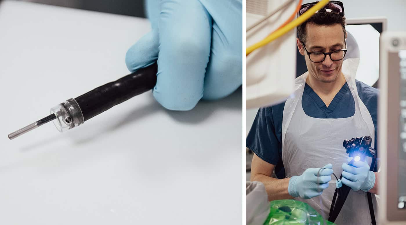
Absolutely, we’ve developed probes that fit inside an endoscope for diagnosing oesophageal cancer.
Earlier in my career I worked on photodynamic therapy. We would look inside the oesophagus with an endoscope to find disease, then give the patient a phototoxic drug that would target the diseased cells. Shining light on the drug causes it to generate singlet oxygen that kills the cancer cells. But I realized that the light we were using could also be used for diagnosis.
Currently, to find this invisible disease, you have to take many, many biopsies. But our in vivo probes allow us to measure the molecular composition of the oesophageal lining using Raman spectroscopy, to be and determine where to take biopsies from. Oesophageal cancer has a really bad outcome once it’s diagnosed symptomatically, but if you can find the disease early you can deliver effective treatments. That’s what we’re trying to do.
Tiny but mighty (left) A Raman probe protruding from the instrument channel of an endoscope. (right) Oliver Old, consultant surgeon, passing the probe down an endoscope for a study led by the University of Exeter, with the University of Bristol and Gloucestershire Hospitals NHS Foundation Trust as partners. (Courtesy: RaPIDE Team)
The very weak Raman signal, however, causes problems. With a microscope, we can use advanced filters to remove the incident laser wavelength. But sending light down an optical fibre generates unwanted signal, and we also need to remove elastically scattered light from the oesophagus. So we had to put a filter on the end of this tiny 2 mm fibre probe. In addition, we don’t want to collect photons that have travelled a long way through the body, so we needed a confocal system. We built a really complex probe, working in collaboration with John Day at the University of Bristol – it took a long time to optimize the optics and the engineering.
Are there options for diagnosing cancer in places that can’t be accessed via an endoscope?
Yes, we have also developed a smart needle probe that’s currently in trials. We are using this to detect lymphomas – the primary cancer in lymph nodes – in the head and neck, under the armpit and in the groin.
If somebody comes forward with lumps in these areas, they usually have a swollen lymph node, which shows that something is wrong. Most often it’s following an infection and the node hasn’t gone back down in size.
This situation usually requires surgical removal of the node to decide whether cancer is present or not. Instead, we can just insert our needle probe and send light in. By examining the scattered light and measuring its fingerprint we can identify if it’s lymphoma. Indeed, we can actually see what type of cancer it is and where it has come from.
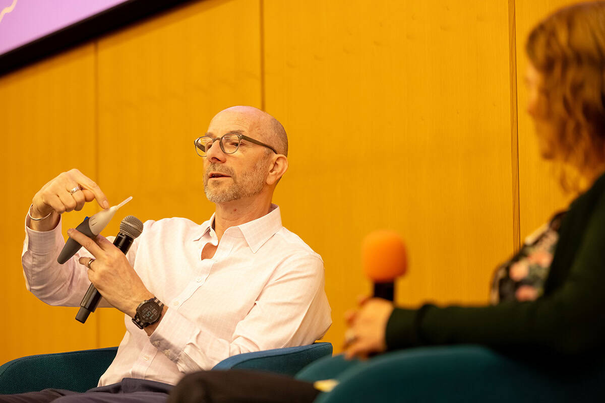
Novel needle Nick Stone demonstrates a prototype Raman needle probe. (Courtesy: Matthew Jones Photography)
Currently, the prototype probe is quite bulky because we are trying to make it low in cost. It has to have a disposable tip, so we can use a new needle each time, and the filters and optics are all in the handpiece.
Are you working on any other projects at the moment?
As people don’t particularly want a needle stuck in them, we are now trying to understand where the photons travel if you just illuminate the body. Red and near-infrared light travel a long way through the body, so we can use near-infrared light to probe photons that have travelled many, many centimetres.
We are doing a study looking at calcifications in a very early breast cancer called ductal carcinoma in situ (DCIS) – it’s a Cancer Research UK Grand Challenge called DCIS PRECISION, and we are just moving on to the in vivo phase.
Calcifications aren’t necessarily a sign of breast cancer – they are mostly benign; but in patients with DCIS, the composition of the calcifications can show how their condition will progress. Mammographic screening is incredibly good at picking up breast cancer, but it’s also incredibly good at detecting calcifications that are not necessarily breast cancer yet. The problem is how to treat these patients, so our aim is to determine whether the calcifications are completely fine or if they require biopsy.
We are using Raman spectroscopy to understand the composition of these calcifications, which are different in patients who are likely to progress onto invasive disease. We can do this in biopsies under a microscope and are now trying to see whether it works using transillumination, where we send near-infrared light through the breast. We could use this to significantly reduce the number of biopsies, or monitor individuals with DCIS over many years.
Light can also be harnessed to treat disease, for example using photodynamic therapy as you mentioned earlier. Another approach is nanoparticle-based photothermal therapy, how does this work?
This is an area I’m really excited about. Nanoscale gold can enhance Raman signals by many orders of magnitude – it’s called surface-enhanced Raman spectroscopy. We can also “label” these nanoparticles by adding functional molecules to their surfaces. We’ve used unlabelled gold nanoparticles to enhance signals from the body and labelled gold to find things.
During that process, we also realized that we can use gold to provide heat. If you shine light on gold at its resonant frequency, it will heat the gold up and can cause cell death. You could easily blow holes in people with a big enough laser and lots of nanoparticles – but we want to do is more subtle. We’re decorating the tiny gold nanoparticles with a label that will tell us their temperature.
By measuring the ratio between Stokes and anti-Stokes scattering signals (which are enhanced by the gold nanoparticles), we can measure the temperature of the gold when it is in the tumour. Then, using light, we can keep the temperature at a suitable level for treatment to optimize the outcome for the patient.
Ideally, we want to use 100 nm gold particles, but that is not something you can simply excrete through the kidneys. So we’ve spent the last five years trying to create nanoconstructs made from 5 nm gold particles that replicate the properties of 100 nm gold, but can be excreted. We haven’t demonstrated this excretion yet, but that’s the process we’re looking at.
This research is part of a project to combine diagnosis and heat treatment into one nanoparticle system – if the Raman spectra indicate cancer, you could then apply light to the nanoparticle to heat and destroy the tumour cells. Can you tell us more about this?
We’ve just completed a five-year programme called Raman Nanotheranostics. The aim is to label our nanoparticles with appropriate antibodies that will help the nanoparticles target different cancer types. This could provide signals that tell us what is or is not present and help decide how to treat the patient.
We have demonstrated the ability to perform treatments in preclinical models, control the temperature and direct the nanoparticles. We haven’t yet achieved a multiplexed approach with all the labels and antibodies that we want. But this is a key step forward and something we’re going to pursue further.
We are also trying to put labels on the gold that will enable us to measure and monitor treatment outcomes. We can use molecules that change in response to pH, or the reactive oxygen species that are present, or other factors. If you want personalized medicine, you need ways to see how the patient reacts to the treatment, how their immune system responds. There’s a whole range of things that will enable us to go beyond just diagnosis and therapy, to actually monitor the treatment and potentially apply a boost if the gold is still there.
Looking to the future, what do you see as the most promising applications of light within healthcare?
Light has always been used for diagnosis: “you look yellow, you’ve got something wrong with your liver”; “you’ve got blue-tinged lips, you must have oxygen depletion”. But it’s getting more and more advanced. I think what’s most encouraging is our ability to measure molecular changes that potentially reveal future outcomes of patients, and individualization of the patient pathway.
But the real breakthrough is what’s on our wrists. We are all walking around with devices that shine light in us – to measure heartbeat, blood oxygenation and so on. There are already Raman spectrometers that sort of size. They’re not good enough for biological measurements yet, but it doesn’t take much of a technology step forward.
I could one day have a chip implanted in my wrist that could do all the things the gold nanoconstructs might do, and my watch could read it out. And this is just Raman – there are a whole host of approaches, such as photoacoustic imaging or optical coherence tomography. Combining different techniques together could provide greater understanding in a much less invasive way than many traditional medical methods. Light will always play a really important role in healthcare.
This article is based on a session at the Institute of Physics’ Celebration of Physics in April 2025.
Dynamic PET imaging is an important preclinical research tool used to visualize real-time functional information in a living animal. Currently, however, the temporal resolution of small-animal PET scanners is on the order of seconds, which is too slow to image blood flow in the heart or track the brain’s neuronal activity. To remedy this, the Imaging Physics Group at the National Institutes for Quantum Science and Technology (QST) in Japan has developed an ultrasensitive small-animal PET scanner that enables sub-second dynamic imaging of a rat.
The limited temporal resolution of conventional preclinical PET scanners stems from their low sensitivity (around 10%), caused by relatively thin detection crystals (10 mm) and a short axial field-of-view (FOV). Thus the QST team built a system based on four-layer, depth-encoding detectors with a total thickness of 30 mm. The scanner has a 325.6 mm-long axial FOV, providing total-body coverage without any bed movement, while a small inner diameter of 155 mm further increases detection efficiency.
“The main application of the total-body small-animal PET (TBS-PET) scanner will be assessment of new radiopharmaceuticals, especially for cardiovascular and neurodegenerative diseases, by providing total-body rodent PET images with sub-second temporal resolution,” first author Han Gyu Kang tells Physics World. “In addition, the scanner will be used for in-beam PET imaging, and single-cell tracking, where ultrahigh sensitivity is required.”
Performance evaluation
The TBS-PET scanner contains six detector rings, each incorporating 10 depth-of-interaction (DOI) detectors. Each DOI detector comprises a four-layer zirconium-doped gadolinium oxyorthosilicate (GSOZ) crystal array (16×16 crystals per layer) and an array of multi-anode photomultiplier tubes. The team selected GSOZ crystals because they have no intrinsic radiation signal, thus enabling low activity PET imaging.
The researchers performed a series of tests to characterize the scanner performance. Measurements of a 68Ge line source at the centre of the FOV showed that the TBS-PET had an energy resolution of 18.4% and a coincidence timing resolution of 7.9 ns.
Imaging a NEMA 22Na point source revealed a peak sensitivity of 45.0% in the 250–750 keV energy window – more than four times that of commercial or laboratory small-animal PET scanners. The system exhibited a uniform spatial resolution of around 2.6 mm across the FOV, thanks to the four-layer DOI information, which effectively reduced the parallax error.
In vivo imaging
Kang and colleagues next obtained in vivo total-body PET images of healthy rats using a single bed position. Static imaging using Na18F and 18F-FDG tracers clearly visualized bone structures and glucose metabolism, respectively, of the entire rat body.
Moving to dynamic imaging, the researchers injected an 18F-FDG bolus into the tail vein of an anesthetized rat for 15 s, followed by a saline injection 15 s after injection. They acquired early-phase dynamic PET data every second until 27 s after injection. To enable sub-second PET imaging, they used custom-written software to subdivide the list-mode data (1 s time frame) into time frames of 0.5 s, 0.25 s and 0.1 s.
Dynamic PET images with a 0.5 s time frame clearly visualized the blood stream from the tail to the heart through the iliac vein and inferior vena cava for the first 2 s, after which the tracer reached the right atrium and right ventricle. At 4.0 s after injection, blood flowed from the left ventricle into the brain via the carotid arteries. The cortex and kidneys were identified 5.5 s after injection. After roughly 17.5 s, the saline peak could be identified in the time-activity curves (TACs).
At 0.25 s temporal resolution, the early-phase images visualized the first pass blood circulation of the rat heart, showing the 18F-FDG bolus flowing from the inferior vena cava to the right ventricle from 2.25 s. The tracer next circulated to the lungs via the pulmonary artery from 2.5 s, and then flowed to the left ventricle from 3.75 s.
The TACs clearly visualized the time dispersion between the right and left ventricles (1.25 s). This value can change for animals with cardiac disease, and the team plans to explore the benefit of fast temporal resolution PET for diagnosing cardiovascular and neurodegenerative diseases.
The researchers conclude that the TBS-PET scanner enables dynamic imaging with a nearly real-time frame rate, visualizing cardiac function and pulmonary circulation of a rat with 0.25 s temporal resolution, a feat that is not possible with conventional small-animal PET scanners.
“One drawback of the TBS-PET scanner is the relatively low spatial resolution of around 2.6 mm, which is limited by the relatively large crystal pitch of 2.85 mm,” says Kang. “To solve this issue, we are now developing a new small-animal PET scanner employing three-layer depth-encoding detectors with 0.8 mm crystal pitch, towards our final goal of sub-millimetre and sub-second temporal resolution PET imaging in rodent models.”
Japanese company ispace says it believes its second lunar lander mission crashed because of problems with a laser rangefinder used to determine altitude during its descent.
BIFROST is a Danish-Swedish surveillance satellite originally developed by Space Inventor. Since then, Terma, Gatehouse SatCom, DTU, and Swedish Unibap Space Solutions have joined the development project, which is financially […]
A consortium of Ukrainian, Nordic and eastern European companies is hoping to raise more than 100 million euros ($115 million) to build a constellation of 70-plus imagery satellites that would provide intelligence along Russia’s border.
Learn more about the new study which suggests humans had to adapt to different environments within the African continent before being able to successfully migrate out of it.
The versatile cannabis plant could, some scientists think, one day be useful for lunar and Martian colonists. For now, researchers will subject its seeds to radiation in orbit and see what happens.
The Golden Dome initiative represents the largest missile defense effort in United States history — a $175 billion shield against weapons of mass destruction, aiming to be fully operational within […]