Nanoparticles demonstrate new and unexpected mechanism of coronavirus disinfection
The COVID-19 pandemic provided a driving force for researchers to seek out new disinfection methods that could tackle future viral outbreaks. One promising approach relies on the use of nanoparticles, with several metal and metal oxide nanoparticles showing anti-viral activity against SARS-CoV-2, the virus that causes COVID-19. With this in mind, researchers from Sweden and Estonia investigated the effect of such nanoparticles on two different virus types.
Aiming to elucidate the nanoparticles’ mode of action, they discovered a previously unknown antiviral mechanism, reporting their findings in Nanoscale.
The researchers – from the Swedish University of Agricultural Sciences (SLU) and the University of Tartu – examined triethanolamine terminated titania (TATT) nanoparticles, spherical 3.5-nm diameter titanium dioxide (titania) particles that are expected to interact strongly with viral surface proteins.
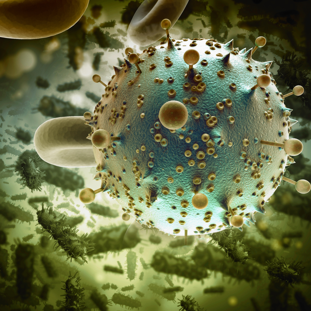
They tested the antiviral activity of the TATT nanoparticles against two types of virus: swine transmissible gastroenteritis virus (TGEV) – an enveloped coronavirus that’s surrounded by a phospholipid membrane and transmembrane proteins; and the non-enveloped encephalomyocarditis virus (EMCV), which does not have a phospholipid membrane. SARS-CoV-2 has a similar structure to TGEV: an enveloped virus with an outer lipid membrane and three proteins forming the surface.
“We collaborated with the University of Tartu in studies of antiviral materials,” explains lead author Vadim Kessler from SLU. “They had found strong activity from cerium dioxide nanoparticles, which acted as oxidants for membrane destruction. In our own studies, we saw that TATT formed appreciably stable complexes with viral proteins, so we could expect potentially much higher activity at lower concentration.”
In this latest investigation, the team aimed to determine whether one of these potential mechanisms – blocking of surface proteins, or membrane disruption via oxidation by nanoparticle-generated reactive oxygen species – is the likely cause of TATT’s antiviral activity. The first of these effects usually occurs at low (nanomolar to micromolar) nanoparticle concentrations, the latter at higher (millimolar) concentrations.
Mode of action
To assess the nanoparticle’s antiviral activity, the researchers exposed viral suspensions to colloidal TATT solutions for 1 h, at room temperature and in the dark (without UV illumination). For comparison, they repeated the process with silicotungstate polyoxometalate (POM) nanoparticles, which are not able to bind strongly to cell membranes.
The nanoparticle-exposed viruses were then used to infect cells and the resulting cell viability served as a measure of the virus infectivity. The team note that the nanoparticles alone showed no cytotoxicity against the host cells.
Measuring viral infectivity after nanoparticle exposure revealed that POM nanoparticles did not exhibit antiviral effects on either virus, even at relatively high concentrations of 1.25 mM. TATT nanoparticles, on the other hand, showed significant antiviral activity against the enveloped TGEV virus at concentrations starting from 0.125 mM, but did not affect the non-enveloped EMCV virus.
Based on previous evidence that TATT nanoparticles interact strongly with proteins in darkness, the researchers expected to see antiviral activity at a nanomolar level. But the finding that TATT activity only occurred at millimolar concentrations, and only affected the enveloped virus, suggests that the antiviral effect is not due to blocking of surface proteins. And as titania is not oxidative in darkness, the team propose that the antiviral effect is actually due to direct complexation of nanoparticles with membrane phospholipids – a mode of antiviral action not previously considered.
“Typical nanoparticle concentrations required for effects on membrane proteins correspond to the protein content on the virus surface. With a 1:1 complex, we would need maximum nanomolar concentrations,” Kessler explains. “We saw an effect at about 1 mM/l, which is far higher. This was the indication for us that the effect was on the whole of membrane.”
Verifying the membrane effect
To corroborate their hypothesis, the researchers examined the leakage of dye-labelled RNA from the TGEV coronavirus after 1 h exposure to nanoparticles. The fluorescence signal from the dye showed that TATT-treated TGEV released significantly more RNA than non-exposed virus, attributed to the nanoparticles disrupting the virus’s phospholipid membrane.
Finally, the team studied the interactions between TATT nanoparticles and two model phospholipid compounds. Both molecules formed strong complexes with TATT nanoparticles, while their interaction with POM nanoparticles was weak. This additional verification led the researchers to conclude that the antiviral effect of TATT in dark conditions is due to direct membrane disruption via complexation of titania nanoparticles with phospholipids.
“To the best of our knowledge, [this] proves a new pathway for metal oxide nanoparticles antiviral action,” they write.
Importantly, the nanoparticles are non-toxic, and work at room temperature without requiring UV illumination – enabling simple and low-cost disinfection methods. “While it was known that disinfection with titania could work in UV light, we showed that no special technical measures are necessary,” says Kessler.
Kessler suggests that the nanoparticles could be used to coat surfaces to destroy enveloped viruses, or in cost-effective filters to decontaminate air or water. “[It should be] possible to easily create antiviral surfaces that don’t require any UV activation just by spraying them with a solution of TATT, or possibly other oxide nanoparticles with an affinity to phosphate, including iron and aluminium oxides in particular,” he tells Physics World.
The post Nanoparticles demonstrate new and unexpected mechanism of coronavirus disinfection appeared first on Physics World.